Welcome
|
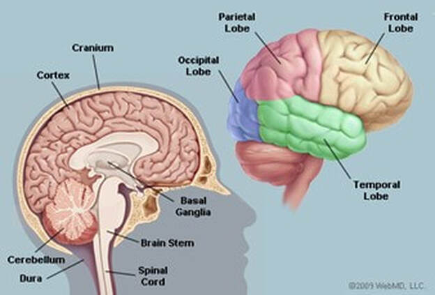
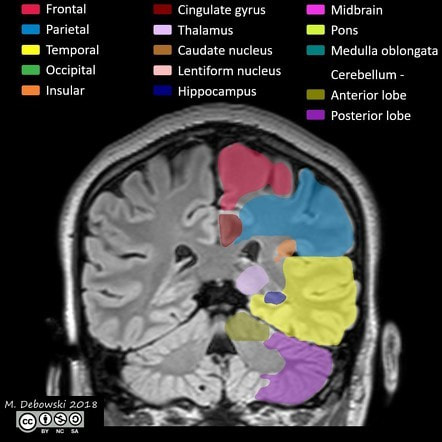
The brain is an organized and structured network with highly complexed functions and thousands of neurons created by Allah (The Most High). Amongst the functions are the six senses: sight, smell, proprioception, touch, taste and hearing covered last month where the brain receives nerve impulses via neurons from the sensory organs. Regardless of the human's technical, knowledge and power, a brain cannot be re-created and this is due to the resistance and signaling mechanisms it encompasses and was described by Penfield as a small universe. The aim of this article is not to provide extensive detail on the anatomy and physiology of the brain but to merely interconnect the verses from the Glorious Quran that associates it with understanding the anatomical-functional of the brain. Allah (The Most High) states: “This is the creation of Allah. So show Me what those other than Him have created. Rather, the wrongdoers are in clear error.” [Quran, Surah Luqman, Verse 11] “And He gave you from all you asked of Him. And if you should count the favor of Allah , you could not enumerate them. Indeed, mankind is [generally] most unjust and ungrateful.” [Quran, Surah Ibrahim, 14:34] “And if you should count the favors of Allah , you could not enumerate them. Indeed, Allah is Forgiving and Merciful.” [Quran, Surah Nahl, 16:18] The malfunction of the neural structure of the brain can cause tumours, injuries and other consequences. Tumours of the brain arise from neoplasms in the meninges and intracranial tissues. The incidence of brain tumours accounts for 2% of all cancers where there are more than 130 types of brain tumours (McKinney, 2004). Tumours can be differentiated based on the histological subtype, grade, stage, biology, presenting symptoms and prognosis and; this influences the type of treatment. The main type of brain tumour is supratentorial that occurs in the frontal, temporal and parietal lobes. Examples of tumours of the glia are glioblastoma, oligodendroblastoma and astrocytoma which increases with age but is less able to be detected in the elderly due to comorbidity conditions, for instance, strokes (Cancer Research UK, 2020). The main modalities of cancer treatment for glioblastomas and lymphoma are radiotherapy, chemotherapy, and surgery (Minniti et al. 2017). However, high doses of ionising radiation decrease the quality of life and increase the risk of neurological toxicity that is secondary to comorbidity (McKinney, 2004). Originally, radiation has been used as a single agent therapy but had little effect on the survival rates. However, an optimal administration of radiotherapy is required for co-adjuvant treatment with chemotherapy, but requires informed clinical decision making and design of randomized trials. This highlights the multidisciplinary approach to create an effective treatment paradigm for patients with brain tumours. Bonner et al. (2020) revealed that silencing of the Microtubule actin crosslinking factor 1 (MACF1), a cytoskeletal protein has anti-tumour properties for patients with glioblastomas where it increases the specificity of radiosensitization, decreasing the viability and migration of glioblastoma cells and impairing translational regulatory processes. There are two types of brain tumours: primary and secondary. Primary tumours of the central nervous system (CNS) initiate in any area of the brain where it can be benign or malignant (LaPointe et al. 2018). Benign tumours are within a defined area and; patients are less likely to have a remission following treatment; whereas malignant tumours can progress to other areas of the brain. Each year, 12100 primary tumours are diagnosed, and this includes the spinal cord, meninges and neurones (nerves) leading to the brain (Cancer Research UK, 2020). Tumours of the brain and spinal cords affect adult and children and; it is the second most common type of paediatric tumours in the UK but is more common in the elderly where it is estimated that for every 100 patients who are 75 or older, 25 people are diagnosed with brain or spinal tumours (Cancer Research UK, 2020). Secondary tumours grow faster and are much difficult to treat with a 5-year overall survival rate (LaPointe et al. 2018). According to statistics, there are 5500 people in the UK diagnosed with malignant cancers (Cancer Research UK, 2020). Gliomas are the most common malignant tumours that occur in adults (LaPointe et al. 2018). The introduction of molecular and genetic markers in the health service has improved prognosis and characterisation of tumours more accurately. Boudreau et al. (2005) revealed that genetic alterations, for instance, IDH, 1p/19q codeletion, RELA-fusion and H3 Lys27Met can be combined with histological techniques in the WHO classification of CNS tumours which aids in the understanding of the diagnosis and treatment of brain tumours. Advanced techniques, for instance, cytological staining and histopathological technology can provide a basis to understand neuroanatomy and increase the specificity of tumour diagnosis. Radiological diagnostic techniques, for instance, positron emission tomography (PET) measures the cerebral blood flow whereas, functional magnetic resonance imaging (MRI) and computed tomography (CT) can be used to understand the structure and function of the brain in response to the cognitive, motor and sensory tasks where the regional and laminar distribution of the human cerebral cortex can be observed (Marshall and Morriss-Kay, 2004). This is needed to complement with an understanding of the model systems to increase detection rates, improve the differential diagnosis of brain tumours that have previously been differentiated as stroke or metastatic tumours (McKinney, 2004). The delivery of care to cancer patients is also influenced by geographical differences, in the UK, cancer treatment is more advanced, and patients have long-term survival. However, the survival rate of brain tumours is low in comparison to other cancers and; there are also differences in its subtypes. For instance, meningiomas, have a better prognosis whereas; patients with glioblastoma multiforme (GBM) have the poorest survival in all age groups. The causes of brain tumours One of the risk factors of brain tumours is the accumulation of genetic alterations caused by chemical, physical and biological factors that evade regulatory mechanisms of neuronal cells and destruct the immune system. Genetic predisposition, for instance, tuberculosis, neurofibromatosis 1 and 2 and nevoid basal cell carcinoma syndrome and mutations of the Guardian of the Genome, p53 gene that can cause Li Fraumeni syndrome has been linked with gliomas (McKinney, 2004). It has also been reported that genes involved in cellular metabolic processes, for instance, DNA stability and repair, detoxification and oxidation can cause brain tumours (McKinney, 2004). Environmental carcinogens such as transplacental N-nitroso compounds (ethyl and methyl nitrosourea) that can cross the blood-brain barrier can have a dual effect. Dietary and environmental N-Nitroso compounds can induce tumours and can have an anti-oxidant effect where it can be found in fruits and vegetables. (McKinney, 2004). However, there are inconsistent reports made on the association between the constituent of the tobacco smoke, hair products, pesticides, pollution with the incidence of tumours and solvents (McKinney, 2004). This requires further research. The role of prokaryotes such as retrovirus and adenovirus have different putative roles causing brain tumours and is caused by the difference in the viral DNA sequences (McKinney, 2004). However, other viruses that cause influenza and varicella (chickenpox) have been linked with brain tumours but; this varies with the seasonality (McKinney, 2004). This suggests that the role of immunological factors in infections and the causation of brain tumours requires further research (LaPointe et al. 2018). The anatomy and physiology of the brain. To gain an accurate understanding of the anatomical-functional perspective of the brain, neuroimaging, electrophysical and; lesion studies have been conducted. The brain forms part of the central nervous system alongside the spinal cord and cranial nerves. The peripheral system consists of spinal nerves that stem from the spinal cord and the autonomous nervous system. The brain is a complex organ that comprise billions of organs and whose weight differs between a man and woman where it is 1336 grams and 1198 grams respectively, however, the difference in weight has no impact on intelligence nor function (Jawabli and Sharma, 2019). There are three fundamental areas in the brain: cerebrum, cerebellum and the brainstem with defined and precise functions as illustrated in Figure 1. The soul dominates the brain and; the soul understands the signals transmitted. Allah (The Most High) states: “Allah is˺ the One to Whom belongs the kingdom of the heavens and the earth, Who has never had ˹any˺ offspring, nor does He have a partner in ˹governing˺ the kingdom. He has created everything, ordaining it precisely.” [Quran, Surah Al-Furqan, 25:2]. The cerebrum contains the main portion of the brain and consists of two cerebral hemispheres. The longitudinal fissure divides the left and right hemispheres, whereas; the corpus callosum connects the lower area of the brain. The brain is composed of two types of cells: neurons and glial cells where the former transmits nerve impulses or signals whereas glial cells are non-neuronal cells that provide support in transmitting the signals from one neurone to another. It contains a myelin sheath that provides insulation. Other functions are maintaining homeostasis and nutrition (Baum et al. 2020). Together, the glial cells, neurons and; the cerebrum forms the human cerebral cortex. Allah (The Most High) states: So which of the favours of your Lord would you deny? [Quran, Surah Al Rahman, 55: 37-38] The cerebral cortex is the outer layer of the cerebrum that extends from the unimodal sensory cortex to the transmodal association cortex (Baum et al. 2020). It consists of grey matter and is fundamental for consciousness and neuronal computations, for instance, voluntary movement memory, communication, thought and; perception (Marguelis et al. 2016). It is anchored in the lower area of the brain by the white matter that comprises of fibres between neurons whose function is to coordinate neural activity (Baum et al. 2020). Additional features of the cerebral cortex consist of grooves of various sizes that are separated by bulges called gyri. This is where nerve impulses move from one gyrus to another and lobe to lobe. Small grooves are called sulci and the larger grooves are called fissures (Baum et al. 2020). Figure 1: Annotation of the brain (Hoffmann, 2019) The cerebrum is divided further into lobes where each cerebral hemisphere consists of frontal, temporal, parietal and occipital that have a specialised function that complements one another. Therefore, malfunction or injury or a tumour arising can influence the ability to communicate. For instance, if one cannot speak, then it will influence their ability to read. Transmission of nerve impulses influences the motor cortex, where the left side would cause the right side of the body to move (Gilbert-Jaramillo et al. 2019). The frontal lobe contains five neural centres and is situated in the pre-frontal cortex behind the forehead. It is required to think, make sound decisions, memory, use of initiative, character, personality, behaviour, and truthfulness (Gray, Warwick and Williams, 1980; Jawabri and Sharma, 2016). There are different types of memories in which the brain processes and in the case of the frontal lobe, it focuses on the prospective memory which involves planning daily to long-term goals (Neulinger et al., 2016). The ability to make sound decisions involves the three aspects: learning, reasoning and creativity (Collins and Koechlin, 2012). The first neural centre in the frontal lobe is the motor speech area of Broca situated in the posterior inferior frontal gyrus (Flinker et al. 2015). It functions in the production and coordination of voices alongside the pharynx, larynx, tongue, and face (Baum et al. 2020). The primary motor area is situated in the posterior area to the precentral sulcus where it controls the voluntary muscle movement of the left side of the body whereas, the secondary motor area controls the voluntary muscle movement of the right side of the body (Chouinard and Paus, 2006). The frontal eye fields control the movement of the eyes. The fifth function is the frontal area that can control and direct evil actions, lying and deteriorate moral principles that are transmitted from the sensory organs to the effectors. The same verse from the Glorious Quran is reflected upon again where Allah (The Most High) states: So which of the favours of your Lord would you deny? [Quran, Surah Al Rahman, 55: 37-38] Yang et al. (2007) revealed that the pre-frontal cortex contains white matter where there is ca. 23-36% orbital frontal middle and inferior gyri but not superior and frontal gyri present in people who live than the normal controls. The rate of white matter is increased in those who pathologically lie (Yang et al. 2007). However, this has already been stated in the Glorious Quran by Allah (The Most High) where He states: “No! If he does not desist, We will surely drag him by the forelock - A lying, sinning forelock.” [Quran, Surah Al Alaq, 96:15-16] Scholars such as Keith Moore in a conference in 1980 confirmed that the frontal lobe of the brain functions in making informed decisions and direct human behaviour. The fronted bone in the skull protects the frontal lobe/forelock, direct human behaviour and make decisions. In a hadith, Prophet (peace be upon him) mentioned a supplication (dua) about the forelock, He said: There is no-one who is afflicted by distress and grief and says (the above), but Allah will take away his distress and grief, and replace it with joy.” He was asked: “O Messenger of Allah, should we learn this?” He said: “Of course; everyone who hears it should learn it: Oh Allah, I am Your servant, the son of Your servant, the son of your maid-servant, and entirely at Your service. You hold me by my forelock. Your Decree is what controls me, and Your Commands to me are just. I beseech You by every one of Your Names, those which You use to refer to Yourself, or have revealed in Your Book, or have taught to any one of Your creation, or have chosen to keep hidden with You in the Unseen, to make the Qur’an Al-Karim the springtime of my heart, the light of my eyes, the departure of my grief, and the vanishing of my affliction and my sorrow. [Ahmad 1:391, Ibn Hibban] This highlights the importance of the frontal lobe in guiding behaviour and is accountable for the deeds of man and; this is where we prostrate (sujood). Allah (The Most High) states: “Recite, [O Muhammad], what has been revealed to you of the Book and establish prayer. Indeed, prayer prohibits immorality and wrongdoing, and the remembrance of Allah is greater. And Allah knows that which you do.” [Quran, Surah Anqabut, 29:45] The cerebellum is the largest area of the hindbrain and approximately weighs 150g (Singh, 2020). It is separated by the cerebrum by a structure called the tentorium and is situated at the back of the brain below the occipital lobe. The cerebellum consists of functional gradients from the primary motor area to the transmodal regions. The other principal axis initiates from task unfocuses to task-focused regions (Guell et al. 2018). The main function of the cerebellum is to maintain the balance of the voluntary movements of the muscles and limbs, strength, and the contraction force (Singh, 2020). The nerve impulses are sent from the cerebral cortex to the spinal cord via the brainstem whereas; the cerebellum connects with the brainstem via the cerebellar peduncles (Singh, 2020). Other functions of the cerebellum include muscle tone and posture (Singh, 2020). The brainstem is situated in the lower extension of the brain, in front of the cerebellum and is connected to the spinal cord as presented in the MRI scan of the brain in Figure 2. There are three structures present in the brainstem and consists of grey and white matter: the pons (metencephalon), midbrain (mesencephalon), and the medulla oblongata (myelencephalon). The brainstem predominantly regulates the central nervous, respiratory and cardiovascular systems as well as the sleep cycle (Siacca et al. 2019). An additional function of the brainstem is to control consciousness by the reticular activating system. The function of the medulla oblongata is to monitor breathing, blood circulation, and heart rhythm. The midbrain and the pons functions in the ocular motion where the pons has additional functions such as facial sensation and movement, hearing and balance. This is led by the twelve cranial nerves that have specific functions: eye movement (oculomotor, trochlear, abducens), ability to see (optic), movement of the neck and shoulder muscles (accessory), taste sensations (glossopharyngeal, vagus), tongue movement (hypoglossal), hearing and balance (vestibular), smell (olfactory), facial sensation (trigeminal) and the facial nerve for expression, sensation of taste and closing of eyelids (Siacca et al. 2019). Injury or destruction of the cerebrum or the cerebellum can cause the patient to live a vegetative state whereas injury to the brain stem can cause brain death and this is due to its association with the heart and other organs. According to contemporary scholars such as Nadwi (2012), if the cerebrum has died and the doctors have confirmed that the brain is dead, then it is permissible to remove the life support even if his or her heart is functioning. Many of the fuqaha (jurists) are in agreement of this. However, concerning the death of the brainstem, there is a difference in opinion amongst contemporary scholars on whether it is considered the end of human life. The heart should not stop beating for the individual to be deemed dead. This was stated in the Islamic Fiqh Council Conference in 1986 in Amman. From the Shariah, it is thought that a person is dead if the following two signs develop: the heartbeat and breathing are ceased, and doctors confirm cessation cannot be reversed. Secondly, the brain function is ceased completely, and this has been confirmed by three neurologists, that the cessation cannot be reversed and the brain is disintegrated (Ghuddah, 1997). The respected scholars have utilised the people of the Cave as an example of how a loss of sensation and consciousness is insufficient to suggest that one is dead as Allah (The Most High) stated in the following verses: “So We cast [a cover of sleep] over their ears within the cave for several years. [Quran, Surah al-Kahf 18:11] Amongst the respected scholars is Al-Shawkani who explained in his book Fath Al-Qadeer about the above Quranic verse that the ears are shut with sleep to prevent them from hearing sounds (Phillips, 2004). His exegesis is based on Al – Mathoor is otherwise known as Al – Riwayah (Gnomic) and Al-Raei are also known as Al-Dirayah (Opinion) to provide this sound reasoning. Other scholars, for instance, Al-Qurtubi informed that the ears are used as a metaphor to elaborate on the corruption of oversleeping and via the ears, sleep was halted (Phillips, 2004). The ‘number of years’ has been confirmed in the same Surah: “And they remained in their cave for three hundred years and exceeded by nine.” [Quran, Surah Al-Kahf, 18:25] Allah (The Most High) states in the following verse: Then We awakened them that We might show which of the two factions was most precise in calculating what [extent] they had remained in time.” [Quran, Surah al-Kahf 18:12] Al-Qurtubi (2013) (may Allah have mercy upon him) explained that Allah wanted to express His knowledge where he already knew which of the two groups was able to calculate the accurate time (Phillips, 2004). Another respected scholar, Al-Shanqiti (2001) (may Allah have mercy upon him) agrees with this and adds that He (Allah) wanted to clarify to people who amongst the two groups were correct about the length of time but, he did not explain anything about the two groups. Majority of the scholars in Tafseer (exegesis) agree that one group is the People of the Cave, whereas the other group of people were the people of the city in which the youths lived. Other scholars agree that it was about the people who are amongst the believers and those who are amongst the disbelievers (Phillips, 2004). The difference in opinion further highlights the wisdom of the Almighty (The Most High) where one would question and also in how resurrection is real and where we will all be resurrected and; Allah (The Most High) can do all things. It was a test for the youths as they were unaware of how long they had slept. Allah (The Most High) answered their prayers and kept those people away whilst they slept in the cave. It is also a reminder of how we should put trust in Allah (The Most High) every time a trial rises. The state of the youth is concluded in the following verse: “And you would have thought them awake while they were asleep and I turned them on their right sides and their left sides and their dog lay with its front legs stretched out at the entrance of the cave. Had you seen them you would have turned and fled filled with fear of them.” [Quran, Surah al-Kahf 18:18]. This suggests that when the People of the Cave woke up, it was not typically as that of the youth who sleep and wake up relaxed. Some of the scholars suggested that their appearance of being awake was concerning their eyes were open when asleep. Other scholars are in the opinion that they were frequently turning on their right and left sides and He (Allah) did not mention the word ‘I’ because they turned as involuntary body movement. Furthermore, Prophet (peace be upon him) discouraged sleeping on the stomach (Phillips, 2004). There is a discrepancy in the number of times amongst the scholars of Tafseer where Abu Hurairah (may Allah have mercy upon him) mentioned twice a year, others mentioned once a year, Mujahid added it was due every seven years (Al-Qurtubi, 2013; Phillips, 2004; Al-Shanqiti, 2001). The outer brain The brain is surrounded by a bony skull (cranium) whose function is to protect the brain. There are also facial bones. The meninges is situated between the cranium and the brain and is one of the three tissue layers that protect the brain and spinal cord. The innermost layer of the meninges is the pia mater, the middle layer is the arachnoid and the outermost layer being the dura mater respectively. The subdural space divides the dura and arachnoid membrane whereas the subarachnoid space divides the arachnoid and pia and this is where the cerebrospinal fluid flows. In addition, the major arteries of the brain and the arachnoid trabeculae flow in the subarachnoid space. The arachnoid trabeculae are strands of arachnoid mater connective tissue. The epidural space exists between the calvarium in the roof of the skull and the superior surface of the dura (Rea, 2015; Ghannam and Al Kharazei, 2020). The pia mater consists of collagen, reticular fibres and many blood vessels that deepen into the surface of the brain (Rea, 2015). The collagen fibres form bundles outside the reticular tissue that surrounds the spinal cord (Rea, 2015). The outer layer of the pia is the epipial and comprises of collagen fibres whereas; the intima pia layer consists of elastic and reticular fibres (Ghannam and Al Kharazei, 2020). Arachnoid is a delicate, thin, avascular elastic membrane and consists of blood vessels that vary in size. It is situated between the dura and pia mata. The primary function of the arachnoid is involved in the cerebrospinal fluid via the subarachnoid space (Rea, 2015; Ghannam and Al Kharazei, 2020). The dura mater comprises of a bi-layered white non-elastic membrane where the outer layer is known as the periosteum. It comprises of osteoblasts and fibroblasts to give strength to the dura. The meningeal layer is close to the brain tissue. Both layers fuse to form two dural reflections and venous sinuses. The dural reflections consist of two special folds: the tentorium cerebelli and falx cerebri (Rea, 2015). The falx is sickle-shaped and divides the right and left of the cerebral hemispheres whereas the tentorium divides the upper and lower area of the brain where it predominantly runs between the cerebellum and occipital lobes (Rea, 2015). The cerebrospinal fluid (CSF) is a clear fluid in channels that provides support to the brain and spinal cord. The CSF produced in these channels are called ventricles in the choroid plexus; the CSF passes through the subarachnoid via the foramina of Luschka (Rea, 2015). The brain maintains the balance in the production and absorption of the CSF. There are four ventricles in the ventricular system: two lateral ventricles are situated in the cerebral hemispheres that communicate with the third ventricle (thalamus and hypothalamus) via the Foramen of Munro. The third ventricle is situated in the centre of the brain and communicates with the fourth ventricle via the Aqueduct of Sylvius (Rea, 2015). The hypothalamus is a small structure consisting of grey matter and regularly communicates with the pituitary gland to maintain homeostasis. The anterior hypothalamus and preoptic area comprise of temperature-sensitive neurons that respond to fluctuations of the internal temperatures by initiating thermoregulatory responses required to restore a constant temperature (Molitch, 2012). To lower heat production, cutaneous vasodilation, sweating and; panting occurs whereas to elevate the body heat, cutaneous vasoconstriction, shivering and; the production of metabolic heat production occurs (Molitch, 2012). The hypothalamic control is required for several hormonal secretions: vasopressin, oxytocin, thyroid-stimulating hormone (TSH), follicle-stimulating hormone (FSH), adrenocorticotropic hormone (ACTH) and leutinizing hormone (LH). It is also involved in the control of sleep where there are specialised hormones situated in the rostral and caudal area of the hypothalamus responsible for this. This highlights the importance of the hypothalamus in the sleep-wake cycle (Henning, 2001; Card and Rinaman, 2002, Puelles et al. 2012). The hypothalamus also forms part of the emotional Limbic System where it is at the centre with access to various neural pathways. The function of the limbic system is in various types of emotions from positivity (love, joy and confidence) and negativity (sad, anger and anxiety) as well as memory (Adnan, 2012). Besides the hypothalamus, the emotional limbic system also consists of the hippocampus, limbic cortex, septal area, amygdala and the subicular complex (Rajmohan and Mohandas, 2007). The hippocampus is a trilaminate structure that is ca. 5cm in size and consists of the outer, middle pyramidal and inner polymorphic layer (Rajmohan and Mohandas, 2007). The amygdala is an almond-shaped structure within the temporal lobe and consists of various forms of nuclei: basolateral, cortical-like, centromedial, amygdaloid and extended amygdala. The limbic cortex is composed of the cingulate and parahippocampal gyri and extends from the anterior of the amygdala to the splenium of the corpus callosum which comprises of the dentate gyrus, hippocampus and subicular complex (Rajmohan and Mohandas, 2007). The septal area consists of grey matter and, the structure of the limbic system collectively is adapted to perform its function as mentioned in the Quran in verse 25:2. Figure 2: An MRI scan of the brain (Rasuli and Elgendy, 2020) Overall, the brain is a complexed structure and; its anatomy and functional organization allow us to sense, question and respond to matter through behaviour and language or speech. The cerebral cortex primarily plays a key role in this from emotion to cognition, movement and memory and; this is coordinated with the cerebellar cortex. Next month, we will discuss the cardio physiology of the heart and what the Glorious Quran states about it. The human body is filled with signs and greatness of Allah's creativity by the ability to know and understand what is wrong from what is right with a clear mindset that ignites peace, love, harmony amongst humanity and I end this section with the following verse from the Quran where Allah (The Most High) states: So which of the favours of your Lord would you deny? [Quran, Surah Al Rahman, 55: 37-38] References
Adnan, A. (2012) The Uniqueness Of The Brain Creation. Available online: https://knowingallah.com/en/articles/the-uniqueness-of-the-brain-creation/ Al-Qurtubi, M.A. (2013) Al-Jami‘ li Ahkam al-Qur’an, 9 – 10. Beirut: Risalah Publishing Al-Shanqiti, M. A (2001) Adwaa Al-Bayaan, 4. Beirut: Dar ibn Hazm. Al-Shawkani, M.A. (1994) Fath Al-Qadir, 5, Beirut: Dar Al-Fikr. Baum, G., Cui, Z., Roalf, D., Ciric, R., Betzel, R., Larsen, B., Cieslak, M., Cook, P., Xia, C., Moore, T., Ruparel, K., Oathes, D., Alexander-Bloch, A., Shinohara, R., Raznahan, A., Gur, R., Gur, R., Bassett, D. and Satterthwaite, T., (2019) Development of structure–function coupling in human brain networks during youth. Proceedings of the National Academy of Sciences, 117(1), pp.771-778. Bonner, K., Borlay, D., Kutten, O. and Quick, Q., (2020) Inhibition of the Spectraplakin Protein Microtubule Actin Crosslinking Factor 1 Sensitizes Glioblastomas to Radiation. Brain Tumor Research and Treatment, 8(1), p.43. Boudreau, C., Yang, I. and Liau, L., 2005. Gliomas: advances in molecular analysis and characterization. Surgical Neurology, 64(4), pp.286-294. Cancer Research UK (2020) ‘What are Brain Tumours?’ Available online: https://www.cancerresearchuk.org/about-cancer/brain-tumours/about Card, J. and Rinaman, L., (2002) Hypothalamus. Encyclopedia of the Human Brain, pp.525-535. Chouinard, P. and Paus, T., (2006) The Primary Motor and Premotor Areas of the Human Cerebral Cortex. The Neuroscientist, 12(2), pp.143-152. Clark, R., (2017) Current Topics Regarding the Function of the Medial Temporal Lobe Memory System. Behavioral Neuroscience of Learning and Memory, pp.13-42. Collins, A. and Koechlin, E., (2012) Reasoning, Learning, and Creativity: Frontal Lobe Function and Human Decision-Making. PLoS Biology, 10(3), p.e1001293. Flinker, A., Korzeniewska, A., Shestyuk, A., Franaszczuk, P., Dronkers, N., Knight, R. and Crone, N., 2015. Redefining the role of Broca’s area in speech. Proceedings of the National Academy of Sciences, 112(9), pp.2871-2875. Ghannam, J. and Al Kharazi, K. (2020) Neuroanatomy, Cranial Meninges. Available online: https://www.ncbi.nlm.nih.gov/books/NBK539882/#_NBK539882_pubdet_ Ghuddah, A. (1997) Qararat wa-Tawsiyat Majma' al-Fiqh al-Islami. Damascus: Dar Al-Qalam. Gilbert‐Jaramillo, J., Garcez, P., James, W., Molnár, Z. and Clarke, K., (2019) The potential contribution of impaired brain glucose metabolism to congenital Zika syndrome. Journal of Anatomy, 235(3), pp.468-480. Gray, H., Warwick, R. and Williams, P. (1980) Gray’s Anatomy UK: Churchill Livingstone. Guell, X., Schmahmann, J., Gabrieli, J. and Ghosh, S., (2018) Functional gradients of the cerebellum. eLife, 7. Hennig, J., (2001) Hypothalamus. International Encyclopedia of the Social & Behavioral Sciences, pp.7113-7118. Hoffmann, M. (2019) Picture of the brain. Available online: https://www.webmd.com/brain/picture-of-the-brain#1 Islamic Fiqh Council (1986) Organisation of the Islamic Conference. Jordan: Amman. Jawabri, K. and Sharma, S. (2020) Physiology, Cerebral Cortex Functions. Available online: https://www.ncbi.nlm.nih.gov/books/NBK538496/#_NBK538496_pubdet_ Kiernan, J., (2012) Anatomy of the Temporal Lobe. Epilepsy Research and Treatment, 2012, pp.1-12. Lapointe, S., Perry, A. and Butowski, N., (2018) Primary brain tumours in adults. The Lancet, 392(10145), pp.432-446. Margulies, D., Ghosh, S., Goulas, A., Falkiewicz, M., Huntenburg, J., Langs, G., Bezgin, G., Eickhoff, S., Castellanos, F., Petrides, M., Jefferies, E. and Smallwood, J., (2016) Situating the default-mode network along a principal gradient of macroscale cortical organization. Proceedings of the National Academy of Sciences, 113(44), pp.12574-12579. Marshall, J. and Morriss-Kay, G., (2004) Functional anatomy of the human brain. Journal of Anatomy, 205(6), pp.415-415. McKinney, P.A., (2004) Brain tumours: incidence, survival, and aetiology. Journal of Neurology, Neurosurgery & Psychiatry 75:ii12-ii17. Minniti, G., Filippi, A., Osti, M. and Ricardi, U., (2017) Radiation therapy for older patients with brain tumors. Radiation Oncology, 12(101). Molitch, M., (2012) Neuroendocrinology and the Neuroendocrine System. Goldman's Cecil Medicine, pp.1425-1431. Molnár, Z., Clowry, G., Šestan, N., Alzu'bi, A., Bakken, T., Hevner, R., Hüppi, P., Kostović, I., Rakic, P., Anton, E., Edwards, D., Garcez, P., Hoerder‐Suabedissen, A. and Kriegstein, A., (2019) New insights into the development of the human cerebral cortex. Journal of Anatomy, 235(3), pp.432-451. Nadwi, M., Thackray, S. and Ahmed, S. (2012) Al-Fiqh Al-Islami: According to the Hanafi Madhhab: Zakah, Fasting and Hajj Volume 2 UK: Angelwing Media. Neulinger, K., Oram, J., Tinson, H., O’Gorman, J. and Shum, D., (2015) Prospective memory and frontal lobe function. Aging, Neuropsychology, and Cognition, 23(2), pp.171-183. Nourski, K., (2017) Auditory processing in the human cortex: An intracranial electrophysiology perspective. Laryngoscope Investigative Otolaryngology, 2(4), pp.147-156. Phillips, A.B. (2004) Tafseer Soorah Al-Kahf Qatar: IOU University Press. RajMohan, V. and Mohandas, E., (2007) The limbic system. Indian Journal of Psychiatry, 49(2), p.132. Rasuli, B. and Elgendy, A. (2020) Limbic system. Available online: https://radiopaedia.org/articles/limbic-system?lang=us Rea, P., (2015) Essential Anatomy and Function of the Spinal Cord. Essential Clinical Anatomy of the Nervous System, pp.121-131. Sciacca, S., Lynch, J., Davagnanam, I. and Barker, R., (2019) Midbrain, Pons, and Medulla: Anatomy and Syndromes. RadioGraphics, 39(4), pp.1110-1125. Singh, R. (2020) Cerebellum: Its Anatomy, Functions and Diseases https://www.intechopen.com/online-first/cerebellum-its-anatomy-functions-and-diseases Watson, C., Paxinos, G. and Puelles, L. (2012) The Mouse Nervous System. Available online: https://www.sciencedirect.com/book/9780123694973/the-mouse-nervous-system#book-info Yang, Y., Raine, A., Narr, K., Lencz, T., LaCasse, L., Colletti, P. and Toga, A., (2007) Localisation of increased prefrontal white matter in pathological liars. British Journal of Psychiatry, 190(2), pp.174-175.
0 Comments
Leave a Reply. |
Archives
February 2021
Categories
All
|
© COPYRIGHT 2024. ALL RIGHTS RESERVED.
Proudly powered by Weebly


 RSS Feed