Welcome
|
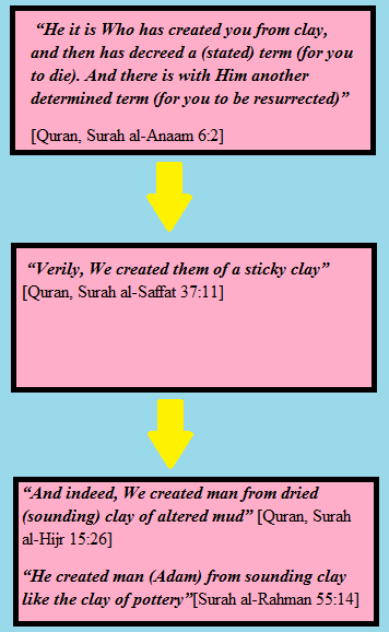
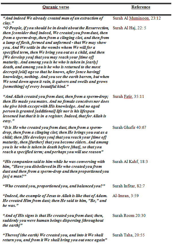
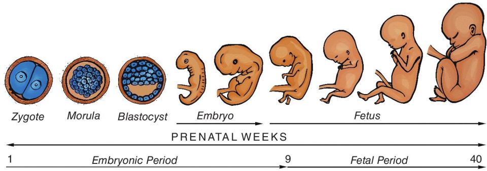
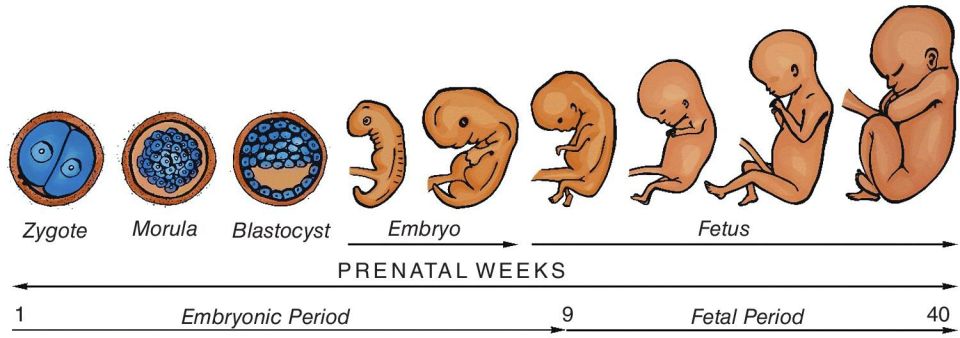
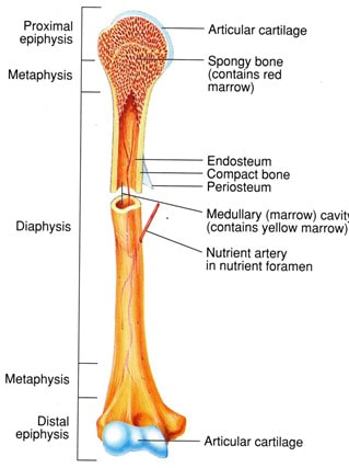
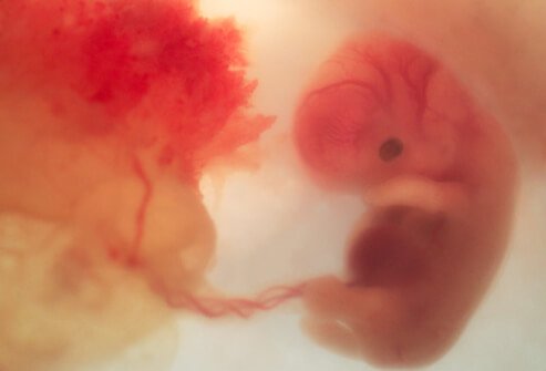
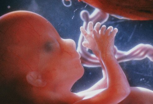
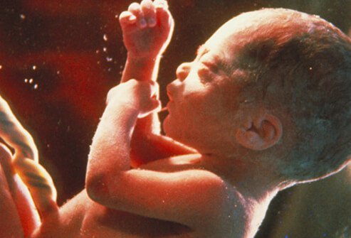
The origin of human nature and how it developed is known as embryology. With today’s advanced imaging and analytical techniques and well-established genetic and experimental models, the development of invertebrates and vertebrates can be studied. The function of this section of my qualitative research review is to prescribe an understanding of how a human is traditionally reproduced and developed from a medical, scientific and Islamic perspective. The Quran and Hadith contain a multitude of evidence on developmental biology; which is a guide for the creation by the Creator, Allah (The Most High) who created man in the best possible manner. “Verily, We created man in the best statue (mould)” [Quran, Surah Tin, 95:4] However, the accuracy and thorough understanding of the Quranic verses and Hadith on the embryonic development were raised after significant discoveries from the ancient times to date have been made by many philosophers and scientists in explaining the key stages in how gametes formed to the full-term pregnancy (World Muslim League, 2000). This ultimately challenges any academic or expert in the field of embryology with such transparency and vivid description of the intrauterine life at the time of the Quran’s revelation; the seventh century where no technology existed. The creation and structure of the human body imply the infinite love of Allah (The Most High) for His creation (Hossain, 2018). Amongst the first philosophers who had an interest in embryology is Aristotle who utilized animal models, for instance, fish, snakes, turtles and insects to perform anatomical studies to discover the development of a human (Saadat, 2009). He compiled it in his book: ‘On the Generation of Animals’ ‘De generatione animalium’ (Boylan, 1984). He also revealed that the foetus is composed of menstrual blood formed in the uterus and is minuscule in the ovum or sperm. However, Allah stated in the following verse: “Was he not a drop of germinal fluid emitted?” [Quran, Surah Al-Qiyamah, 75:37] One of the prominent scholars, Ibn Hajar Al Asqalani (1997) stated that: “Many Anatomists claim that the male fluid has no effect on the creation of the child except for coagulation of blood and that the child is formed from the menstrual blood. However, the Prophet’s Hadith in this chapter refutes this claim”. The findings and conceptions of Aristotle were challenged by Galen because some eggs of models used fertilized, whereas other animals grew spontaneously from meat, for instance, flies (Saadat, 2009). In the second century, Galen described the structure and function of the placenta and the role of placental proteins in embryonic development in his book ‘On the formation of the foetus’ (Longo and Reynolds, 2010). Furthermore, the invention of the microscope by Zacharias Janssen in the sixteenth century which was further developed by Anton van Leewenhoek and Robert Hooke in the seventeenth century who invented the light and the compound microscopes respectively brought wonders of the natural sciences (Wollman et al. 2015; Hooke, 1665). The instruments led to ongoing discoveries and illustrations that contribute to the development of medical and scientific knowledge, particularly in embryology. In 1672, Malphigi studied poultry eggs under the microscope who questioned epigenesist and discovered muscle-forming somites (Keele et al. 1978; Gilbert, 2000). In 1775, Spallanzani revealed that the spermatozoon and ovum are required to fertilize for a human to be developed (Capanna, 1999). This highlight the progression of understanding embryology. Later in the 19th century, Frankie Lillie (1914) discovered the physical and chemical mechanisms in how the fertilization step takes place between the egg and the sperm. Today, researchers utilize various genetic techniques and advanced imaging tools to study the cells. However, the comprehensive methodology of how the human body developed is already present in the Glorious Quran. The Seal of the Prophets, Muhammad (peace be upon him) was illiterate and; he was taught by Allah (The Most High). It was through Prophet Muhammad (peace be upon him), many scholars sought advice on the Tafsir of the Quranic verses to develop their knowledge and understanding of Allah (The Most High) divine wisdom (Moore, 1986). Thus, with certainty, Allah has created human and monitored the reproduction and embryonic development phases until the baby enters Earth. The child then grows and lives and dies as he or she destined to be. Modern technology and knowledge cannot measure and understand the infinite dimensions in which this universe possesses. Allah (The Most High) manages all affairs who created everything with His Mercy, wisdom and intellect. The development of man consists of physical creation, spirit, fitrah and light (Shakir, 2018). The first person that landed on Earth was Adam (peace be upon him) and was physically created by Allah (The Most High) in His image with ‘Both His Hands’ as mentioned in the Tafsir of Ibn Abbas (may Allah have mercy upon him) in the following Quranic verse: “(He said) Allah said to him: (O Iblis!) O wicked one! (What hindereth thee from falling prostrate before that which I have created with both My hands) what prevented you from prostrating before that which I have shaped with My hands? (Art thou too proud) to prostrate before Adam (or art thou of the high) or are you of those who disobey My command?” [Quran, Surah Saad, 38:75] This emphasizes the great care in creating Adam without the intermediary of a mother nor father (Shakir, 2018). Some jurisprudence such as Abu Thawr and Ibn Khuzaymah explained that it was in Adam’s image (Melchert, 2011). The Ruh (spirit) comprises the senses, wisdom and physical stature of the humankind as mentioned in the following Quranic verse: “And when He had made him upright and breathed into him of His spirit” [Quran, Surah Saad, 38:72] In another narration, Allah (The Most High) states: "Thereafter, He moulded him and breathed into him of His Spirit, and He made for you hearing and beholding (s) (i.e., eyesight) and heart-sights; (i.e., perception) little do you thank (Him)." [Quran, Surah Sajdah, 32:9] This indicates that the spirit was breathed into the human body and soul. There are two types of spirits: Al-Sugrah where death occurs during the sleep and Al-Kubrah; the actual death. In each form, the spirit leaves the body but in Al-Sugrah returns (Setiawan et al. 2019). Al-Razi implied that once the spirit has entered the body, it travels to every part and described metaphorically as the air breathed into a vessel. The Fitrah (natural disposition) occurs from Allah (The Most High) and has several perspectives based on its etymology otherwise known as ‘lafaz al mushtarik’ (Rahman, 2012; Kamus Dewan, 1996). Al-Jurjani, (2003) stated that the Fitrah is the divine form created within the child in the womb of the mother where he or she accepts Islam as the religion of the truth by nature. Another understanding of its meaning is the inner consciousness of a person (Bhat, 2016). Ibn Manzur (1988) explained that when Allah blesses someone and grant them peace and teaches man to repeat certain words when lying down to sleep, that if one were to die the same night, one would die upon the truth; the fitrah. “So direct your face toward the religion, inclining to truth. [Adhere to] the fitrah of Allah upon which He has created [all] people. No change should there be in the creation of Allah. That is the correct religion, but most of the people do not know” [Quran, Surah Al Rum, 30:30] Amongst the 99 attributes of Allah (The Most High) is Al-Fatir, the creator and inventor who created the fitrah in man. Light also forms part of the human creation given from Allah (The Most High) as mentioned in the following verse below (Al-Razi, 2010): “On the Day you see the believing men and believing women, their light proceeding before them and on their right, [it will be said], "Your good tidings today are [of] gardens beneath which rivers flow, wherein you will abide eternally." That is what is the great attainment.” [Quran, Surah Hadid, 57:12] Therefore, the people who have affirmed their faith will be recognised by Allah (The Most High) and is based on their righteous deeds, the piety of their characters and this correlates with the luminous and intensity of the light. Ibn Kathir narrated that Abdullah ibn Masood (may Allah have mercy upon him) and collected by Ibn Abu Hatim and Al-Tabari that there will be light running forward before them. "They will pass over the Sirat according to their deeds. Some of them will have a light as large as a mountain, some as a date tree, some as big as a man in the standing position. The least among them has light as big as his index finger, it is lit at times and extinguished at other times.'' Therefore, the development of man consists of the physical creation and other dimensions highlighted. However, the scope of this section is to focus on the physical creation that associates medicine and science with the Glorious Quran. The formation of the human from clay The creation of man has been detailed throughout the Quran more than other creation as presented in Table 1. At first, the dust is mixed with water to form clay that was sticky and viscous. Based upon the chemical composition of clay we would discover various elements in the human body that assemble them such as carbonates, water-soluble salts, minerals such as silicon, calcium and iron (Vakalova et al. 2018). It was informed by Ibn Manzur (2010) that the term Lazeeb is extracted from the term lazooba which means a sticky solid. The sticky product is then converted into muntin stinking. Muntin is thin strips of wood that divide the glass panels. Allah (The Most High) uses the term Al hama which is a form of black mud and has turned to masnoon due to a change in water. When mixing it with sand, it forms a ‘salsal' which is dried clay and this has been agreed by various scholars (Ibn Manzur, 1988; Al-Razi, 2010). Figure 1: The process of the formation of sounding clay Therefore, our Prophet Adam (peace be upon him) was formed from sounding clay that is black, soft mud that has altered. Ibn Jarir, Ibn Qayyim Al Jawziyah and Qatadah Ibn Dia’amah Sadusi Basri and other scholars agree with this. Ibn al-Qayyim Al Jawziyah (1960) said: “When the perfection, complete power, all-encompassing knowledge, ever-executed will and utmost wisdom of the Lord decreed that His creation should be of materials of different kinds and that they should vary in their forms and attributes and natures, His wisdom decreed that He should take a handful of dust from the earth, then mix it with water. So it became like black stinking mud. Then the wind was sent upon it and it dried out until it became clay-like pottery. Then it was given shape and limbs and faculties, and each part of it was given a shape suited to its purpose”. Abu Musa Al-Ashari (may Allah have mercy upon him) who said: “I heard the Messenger of Allah (peace and blessings of Allah be upon him) say: ‘Allah created Adam from a handful that He gathered from the entire earth, so the sons of Adam come to like the earth. Some of them are red, some are white, some are black and some are in between. Some of them are easy, some of them are difficult, some are evil and some are good.” [Al-Tirmidhi, 2955; Abu Dawood, 4693] This evidence provides an insight into the morphogenesis of the materials and architecture in how Allah (The Most High) has created Adam (peace be upon him) in the shape and form He desires. The first human created and generations upon generations initiated. However, human development does not pause at the clay. For Allah (The Most High) states in the Quran: "And indeed We created man (Adam) out of an extract of clay (water and earth); Thereafter We made him (the offspring of Adam) as a Nutfah (mixed drops of the male and female sexual discharge and lodged it) in a safe lodging (womb of the woman); Then We made the Nutfah into a clot (a piece of thick coagulated blood), then We made the clot into a little lump of flesh, then We made out of that little lump of flesh bones, then We clothed the bones with flesh, and then We brought it forth as another creation. So Blessed is Allâh, the Best of creators" [Quran, Surah Al Muminoon 12-14] The transitional changes and time intervals from one developmental phrase from one stage to another are emphasised with total accuracy by Allah (The Most High) by the phrase ‘then' where the clay extract to a drop from the spermatozoon ‘mixes' or fertilises the egg from the ovum to become a zygote (An-Najjar, 2005). At the end of the clot phase, researchers have staged that somites are formed and converts into a Morsel giving the embryo shape. The ‘lump' of flesh form the appendicular and axial skeletons via osteogenesis. The axial skeleton consists of ribs and the cartilaginous vertebral column derived from the sclerotome (Dockter, 1999). The formation of sclerotome and somite chondrogenesis stimulated by two ligand molecules, Noggin and Shh in the notochord and neural tube (Dockter, 1999). The bones are ‘clothed' with flesh (muscle and skin); the dermomyotome. Dermomytome is an epithelial cell layer situated at the dorsal end of the somite which aid in the formation of the dermis and skeletal muscle of the myotome ontogeny and tissues (Ganten et al. 2006). By understanding the morphogenesis, one can understand the underlying processes involved in carcinogenesis, wound healing and organ regeneration (Sieck, 2019; Kahane et al. 2013). Table 1: Some of the Quranic evidence of human created from clay. An insight into the Nutfah (zygote phase) Following the formation of the clay, Nutfah occurs where one male germ cell, spermatozoa required to ejaculate during sexual intercourse to penetrate and fuse with the ovum, the female sex cell to produce a fertilized egg called the zygote. To conceive twins depends on the genetic disposition and environmental factors (Hoekstra et al. 2008). Monozygotic twins are genetically identical and are caused from one zygote and divided into two where they commonly have two amnions, one chorion and one placenta (Hoekstra et al. 2008). On the other hand, dizygotic twins have two zygotes independently fertilized where the outcome consists of two of each of the following: amnions, chorions and placenta where there is ca. 65% of is fused (Hoekstra et al. 2008). Allah (The Most High) described the formation of the zygote in the following verses that a small quantity of both secretions required during the Nutfah phase called Amshaj. The fluid is discharged from both men and women to fertilize in the fallopian tube before descending to the uterus where implantation occurs followed by the development of villosities called Alaq (Sadaat, 2009). William Harvey’s doctrine omne vivum ex ovo translates to all life comes from the egg highlights the importance of the egg and sperm evading the theory of spontaneous generation (Keynes, 1966). Understanding the embryological phases from fertilization to birth through the Quranic verses supplemented with the modern medical and scientific evidence provides us with an anatomical and physiological understanding us to know how we all started (Varga, 2017). “And that He creates the two mates - the male and female. From a sperm-drop when it is emitted” [Quran, Surah Al-Najm, 53:45–46] “Verily, I created humankind from a small quantity of mingled fluids.” [Quran, Surah Insan, 76:2] “And it is He Who has created man from water, and has appointed for him kindred by blood, and kindred by marriage” [Quran, Surah Al Furqan, 25:54] “Then He made his offspring from semen of despised water (male and female sexual discharge)” [Quran, Surah Al-Sajdah, 32:8]. The Amshaj consists of three phases: Khalk, Taqdir and Harth (Sadaat, 2009). Khalk is the fertilization process where the zygote has 46 chromosomes. There are two types of cell division: meiosis and mitosis. Meiosis is derived from the Greek word mitos meaning thread which describes the thread-like appearance of the chromosomes (Albert et al., 2002). It is a regulated process that consists of two successive rounds of cell division to create gametes. Meiosis differs from mitosis in the regulatory mechanisms but is similar in the process of segregation and recombination (O’Connor, 2008). Allah (The Most High) has designed the female and male reproductive systems whose function is to produce these gametes and hormones and support the development of the foetus and delivering it to this enigmatic world (Betts et al., 2013). The female reproductive system situated in the pelvic cavity where the gamete produced is called an oocyte. Oogenesis initiates in the oogonia, where the primordial follicles develop in the primitive ovarian stem cells and; the primary oocytes form via mitosis before birth. They are then arrested during the first meiotic division and then reinitiates during puberty and continues until the menopause stage (Betts et al. 2013). The release of the oocyte signifies the conversion from puberty to a woman known as ovulation. It is estimated that the number of primary oocytes decreases from ca. two million to 400,000 at puberty and then zero post-menopause (Betts et al. 2013; Edwards et al., 1969). “And Allah has made for you from yourselves mates and has made for you from your mates sons and grandchildren and has provided for you from the good things. Then in falsehood do they believe and in the favour of Allah they disbelieve?” [Quran, Surah Al Nahl, 16:72] Figure 2: A schematic presentation of the meiotic process (Marston and Amon, 2005) The second phase of the Amshaj is the formation of facial and other characteristics and is called Taqdir (Sadaat, 2009). The determination of the gender can be identified depending on the type of chromosome that fertilizes the ovum and is facilitated by the cascade of factors induced by the SRY (Sex-determining region of the Y chromosome) (Betts et al. 2013). The male gender has an XY chromosome whereas, a female has an XX chromosome. If the Y chromosome of the man fertilizes the ovum it is a boy and if it is an X chromosome it is a girl. In the female and male embryos, the tissue is bipotential where the cells can differentiate to form female or male gonads (Betts et al. 2013). This causes a series of cascade where functional SRY gene causes the individual to become a male and, when it is not functional it becomes a female to form spermatogonia and oogonia respectively. The bipotential paradigm can be explained with testosterone where Leydig cells secrete the hormone that can directly differentiate into glans penis present in the male reproductive system or the glans clitoris in the female reproductive system. However, without testosterone, cells differentiate into the clitoris (Betts et al., 2013). Moreover, sustentacular cells also secrete testosterone which stimulates the growth of the male duct, Wolffian and degrades the female duct; Mullerian duct; low levels of testosterone causes the production of the Mullerian duct (Betts et al. 2013). This signifies the significant impact that hormones have to mature reproductive system and develop sexual characteristics. Besides testosterone and oestrogen from the gonads, the luteinizing hormone (LH) functions synergistically with follicle-stimulating hormone (FSH) to induce the growth of follicles and ovulation; they are both secreted in the anterior gland (Raju et al. 2013). The Gonadotropin-releasing hormone (GnRH) is secreted from the hypothalamus and determines the secretion of both LH and FSH that function in maturing the gonads (Marques et al. 2018). “God fashioned humans from a clinging entity.” [Quran, Surah Alaq, 96:2] On the other hand, the bipotential mechanism of differentiation of internal structures of the reproductive system is not apparent in all tissues. For instance, the uterus found in the female and the ductus deferens, epididymis and seminal vesicles in males are derived and produced from one of the duct systems of the embryo (Betts et al., 2013). For it to be functionally active, one must develop appropriately whilst the other degrades (Betts et al., 2013). From an Islamic perspective, Allah (The Most High) has informed us that Eve has been created from the rib of Prophet Adam (peace be upon him) and is has caused significant discussions amongst the scholars of the exegesis. However, it is important to state that within the legal theory, Usul Al-Fiqh, there are several laws of language, Quwaid Al-Lughawiyah, for instance, clarity (Al-Faz Wadihah), reference (Turooq Al-Fiqh), pronunciation (lafz) and qualities (sighaat) to understand the basics and phrases present in Quran and Hadith (Al-Shami, 2015; Zulkifli, 2017). In Surah Al Zumr, Allah says ‘He created you from one soul’ and exemplary scholars such as Al-Tabari (2000), Al-Razi (1981), Ibn Asyur (1984), Al-Suyuti and al-Mahalli, (2007), Ibn Kathir (1980) and Ibn Kathir (2014) explains that the one soul is Adam and the mate is Eve. Therefore, Eve was produced from the left rib of Adam whilst he was asleep. However, Ibn Kathir (1980) does not elaborate on how Eve was created as that of Adam from clay to birth which indicates that they undergo the same process of pregnancy. “It is He who created you from one soul and created from it its mate that he might dwell in security with her. And when he covers her, she carries a light burden and continues therein. And when it becomes heavy, they both invoke Allah, their Lord, "If You should give us a good [child], we will surely be among the grateful." [Quran, Surah Al Araf, 7:189] “O mankind, fear your Lord, who created you from one soul and created from it its mate and dispersed from both of them many men and women. And fear Allah, through whom you ask one another, and the wombs. Indeed Allah is ever, over you, an Observer.” [Quran, Surah Al Nisa, 4:1] “He created you from one soul. Then He made from it its mate, and He produced for you from the grazing livestock eight mates. He creates you in the wombs of your mothers, creation after creation, within three darknesses. That is Allah, your Lord; to Him belongs dominion. There is no deity except Him, so how are you averted?” [Quran, Surah Al-Zumr, 39:6] On the other hand, some scholars disagree that Eve was created in this manner. For instance, Al Maraghi (1946) wherein the following verse in Surah Taha: “Thereof (the earth) We created you, and into it We shall return you, and from it, We shall bring you out once again” [Quran, Surah Taha, 20:55] The term we highlight that Adam and Eve created from the same material, earth. Another scholar, Al-Qismi, opposes the idea of Eve produced from Adam’s rib but of human nature (Al-Qismi, 2003). On the contrary, scholars such as Al-Kirmani (1981) emphasises that Eve created from the rib based on the following hadith that graded as sahih in Ale-Shaikh (2010) publication called Al-Kutub Al Sittah that comprises a compilation of six volumes of hadith: Al-Bukhari, Al Nisai, Muslim, Abu Dawood, Tirmidhi and Ibn Majah. Abu Hurairah (may Allah be pleased with him) narrated that Prophet Muhammad (peace be upon him) wherein the upper area of the rib there is crookedness that can is broken if straightened said: “Treat women kindly, for the woman was created from a bent rib, and the most crooked part of the rib is the top part, so treat women kindly.” [Al Bukhari and Muslim] A day after fertilization, the zygote divides (cleavage) into two cells without a cytosol, additional cleavage take place and, this causes a decrease in cell size per cell division. The cells that are dividing are known as blastomeres (Boiaini et al., 2019). The pre-implantation of the human embryo has two phases: the first three days occur in the fallopian tube and four days in the uterus. In total, the pre-implantation period is a week and; the outcome is a blastocyst implanted into the fundus of the uterine wall (Leese, 2010). This is known as Harth (Saadat, 2009). The zygote descends from the fallopian tube (oviduct) by peristalsis and cilia to the uterus where it is implanted in the endometrium by digesting the endometrial cells to a position in the uterine mucosa (Saadat, 2009). Implantation stimulated by the trophectoderm (TE) cells of the blastocyst (Hyun et al. 2020). The hormones, progesterone, oestrogen and human chorionic gonadotropin (hCG) are produced by the differentiated syncytiotrophoblasts to maintain the development of the embryo (Nwabuobi et al. 2017). On the fourth day, a 16 -32 cell conceptus formed towards the uterus where it appears like a solid mass comprising of blastomere within the zona pellucida; a glycoprotein layer around the embryo. This is called the morula as illustrated in Figure 3 and, there are two rounds of asymmetric divisions (Saini and Yamanaka, 2018). At the fourth cell division, the cell number increases from 8 to 16 and in the fifth cell division, 16 – 32 cells (Saini and Yamanaka, 2018). The conceptus that consists of the embryo and the additional membranes increase in cell number and nourished by the uterine milk secretions from the decidual cells of the endometrium; it is also mediated by progesterone (Ing et al., 1989). The endometrial secretion also causes the uterine lining to thicken (Ing et al., 1989). Figure 3: The embryological phases during the embryonic period and foetal period (Pocket Dentistry, 2017) An insight into the Takhliq phase (embryonic phase) Takhliq is organogenesis where differentiation from cells produces rudimentary structures of tissues and organs (Saadat, 2009). This takes place between the third and eighth week of gestation from germ layers: endoderm, ectoderm and mesoderm (Saadat, 2009). This stage extends from the beginning of the third week to the eighth week of the embryonic period: Alaqah (leech-like), Mudghah (somites), Idham (bones) and Laham (muscles) (Saadat, 2009). Alaqa phase There is several meaning of alaqa: leech, blood clot and a suspended substance and be explained as we go through the embryonic phase (Sadaat, 2009; Moore, 1986). On the fifth day, a transition takes place from a morula to blastocyst and is supplemented by a change in the metabolic pathway from pyruvate and lactate to glucose (Kaneko and DePamphilis, 2013). The formation of the blastocyte otherwise, known as blastula is necessary to determine the mammalian ontogeny and surrounded by a fluid-filled cavity called a blastocoel (Denkey, 1969; Kaneko and DePamphilis, 2013). The blastocyst consists of an inner cell mass (ICM) and trophectoderm (TE) (Marikawa and Alarcon, 2009). As the ICM increase in size, the zona pellicuda becomes thinner and hatches away from the zona pellicuda in the following day (Shafei et al., 2017). The ICM has totipotency of differentiating to various types of cells. For instance, the epithelial cells of the placenta, trophoblasts that surround the outer shell is maintained by the Hippo signalling pathway (Saini and Yamanaka, 2018). Both types of trophoblasts, cytotrophoblasts and syncytiotrophoblasts differentiate to form the chorionic membrane that positions the conceptus into the endometrium and form the foetal part of the placenta (Denker, 1969). The maternal part of the placenta-derived from the deepest layer of the endometrium; decidua basalis and the cytotrophoblasts remodel the maternal blood vessels that surround the chorionic villi and form the chorionic sac which extends into the endometrium (Gest, 2000; James, 2014). The mesenchymal cells from the mesoderm villi differentiate into umbilical blood vessels to connect the embryo with the placenta and allow the exchange of nutrients and removal of waste (Niakan et al. 2012). This is why this phase is described as the blood clot and suspended substance whereby blood vessels a formed and the embryo associates with the placenta (Moore, 1986). On the sixth day, the blastula attaches to the endometrium of the uterine wall for two to three weeks until the 25th day where it appears a ‘leech-like structure’ (Sadaat, 2009; Moore, 1986). The embryo is compared to a leech parasite because the embryo derives blood from the decidua. This is similar to the leech with the host (Kuo and Lai, 2018). The invention of the microscope has facilitated medical scientists to examine the morphological structure of the embryo that could not be achieved in the 7th century due to the miniature size of the embryo during the earlier days of pregnancy (Moore, 1986). This correlates with the following verse, where Allah (The Most High) describes the embryo as a leech-like structure: ‘Then We made the sperm-drop into a clinging clot, and We made the clot into a lump [of flesh], and We made [from] the lump, bones, and We covered the bones with flesh; then We developed him into another creation. So blessed is Allah, the best of creators.’ [Quran, Surah Muminoon, 23:14] During the second week, the blastocyst differentiates into layers producing extraembryonic membranes, chorion, amnion, allantois and yolk sac to protect and support the embryo and ensure implantation takes place (Hyun et al., 2020). The ICM produces a two-layered embryonic disc, the upper layer, epiblast, surrounds the amniotic cavity to form an amnion comprising of fluid and maternal plasma. The hypoblast endoderm is present on the ventral end of the disc and forms the yolk sac with the extraembryonic mesoderm (Hafez, 2017). The chorion is a membrane that surrounds the other membranes whereas the allantois is an excretory duct of the embryo that forms as part of the bladder to remove metabolic waste (Muench et al., 2017). The yolk sac and allantois produces the umbilical cord and allows nutrients to reach the embryo via primitive blood circulation (Pansky, 1982). The germ layers formed are endoderm, ectoderm and mesoderm to shape the foetus (Rehman and Muzio, 2020). The bi-laminar germ disk becomes a tri-laminar germ disk in the third week via gastrulation where multipotency rather than totipotency to form an oval shape and primitive streak on the epiblast to proliferate and develop the structures in the embryo (embryology.ch, 2020a). The epiblast produces the ectoderm, the middle layer mesoderm and the endoderm replaces the hypoblast produces the yolk (Pilato, 2003). The endoderm further differentiates to form two tubes: the gastrointestinal and respiratory tube (Gilbert, 2000). The gastrointestinal tube buds several organs: liver, pancreas and the gall bladder, whereas the respiratory tube bifurcates into two tubes (Gilbert, 2000). Both tubes share an anterior structure known as the pharynx where further epithelial structures, for instance, thymus, thyroids, parathyroid glands and tonsils appear (Gilbert, 2000). The process of the development of organs and the central nervous system is called neurulation. Figure 4: The embryo at 4 weeks during the first trimester (Shiel, 2009) Mudghah (Somites) phase. Following the alaqa phase, the development of the somites occurs and the embryo is described as a chewed up morsel (Moore, 1986). This is because of the small size (1 cm) and it can be chewable by teeth (Moore, 1986). It has a corrugated surface where some parts of the somites are formed and other areas are unformed that correspond to the differentiated and undifferentiated tissues (Sadaat, 2009). The primordial synthesis of the internal ears leads to the early creation of the eyes and then the brain (Moore, 1986). Allah (The Most High) has already started in the following Quranic verse of the transition between the alaqa to mughdah: “I fashioned the clinging entity into a chewed lump of flesh and I fashioned the chewed flesh into bones and I clothed the bones with intact flesh.” [Quran, Surah Al Muminoon 23:14] “O People, if you should be in doubt about the Resurrection, then [consider that] indeed, We created you from dust, then from a sperm-drop, then from a clinging clot, and then from a lump of flesh, formed and unformed - that We may show you. And We settle in the wombs whom We will for a specified term, then We bring you out as a child, and then [We develop you] that you may reach your [time of] maturity. And among you is he who is taken in [early] death, and among you is he who is returned to the most decrepit [old] age so that he knows, after [once having] knowledge, nothing. And you see the earth barren, but when We send down upon it rain, it quivers and swells and grows [something] of every beautiful kind.” [Quran, Surah Al-Hajj 22:5] In a hadith, Anas ibn Malik (may Allah have mercy upon him) narrated by the Prophet Muhammad (peace be upon him) said: “Allah has appointed an angel as the caretaker of the womb, and he would say: My Lord, it is now a nutfa or a sperm-drop; my Lord, it is now a clot; my Lord, it has now become a chewed morsel, and when Allah decides to give it a final shape, the angel says: My Lord, would it be male or female or would he be an evil or a good person? What about his livelihood and his age? And it is all written as he is in the womb of his mother.” [Al-Bukhari and Muslim] During the fourth week, the placenta decreases the size of the yolk sac to facilitate the blood vessels and the gametes. The connection between the placenta and the embryo via the umbilical cord is strengthened throughout the gestation period. The umbilical cord is a blood conduit with a helical and tubular structure that surrounds the amnion and is filled with the mesenchyme Wharton’s jelly; a supportive connective tissue (Spurway et al., 2012). It has one umbilical vein and two umbilical arteries but is not fully developed until the 12th week of the gestation (Jarzembowski, 2014; Spurway et al., 2012). The arteries remove deoxygenated blood into chorionic blood vessels and out of the chorionic villi where it filters the blood and exchanges nutrients and oxygenated blood between the foetal and maternal blood (Carlson, 2014; Denker, 1969). There are various mechanisms in how nutrients and drugs are exchanged, for instance, facilitated diffusion, active transport, passive transport and pinocytosis and the placental transfer rate is dependent on the molecular size and the pharmacological properties of the prescribed drugs (Griffiths et al., 2014). The placenta is permeable to lipophilic substances, for instance, antibiotics that move via facilitated diffusion, whereas it is hydrophilic to glucose and other small hydrophilic solutes (Faber and Anderson, 2012; Reynolds, 1998). Iron and amino acids are transferred by active transport (Faber and Anderson, 2012). Anaesthetic drugs and analgesic drugs can cross the placenta (Griffiths et al., 2014; Reynolds, 1998). The neuroectodermal cells that surround the embryo thicken and become a neural plate where tissues either side produces a neural tube (Pleasure et al. 2014). The neural tube transforms into a rod-shaped notochord and is then altered to a nucleus polposis of discs to facilitate in the generation of neurons (nerve cells) and neuroglial (supportive cells) via neuroepithelial progenitors (Pleasure et al. 2014). The two types of the nervous system, central nervous system (CNS) Aand the peripheral nervous system (PNS) are formed in the embryo at 18 days (Sadaat, 2009). Adhesion molecules, for instance, N-cadherin and neural cell adhesion molecule (NCAM) are transcribed to facilitate the development of the neural tube structure. Folate (Vitamin B12) is also essential for the neural tube where defects such as spina bifida and in severe cases anencephaly can occur in response to its deficiency (Molley et al. 2009). During the fifth week, the size of the neural tube increases at the anterior end and produces three primary vesicles of the brain: prosencephalon (forebrain), mesencephalon (midbrain) and rhombencephalon (hindbrain). The optic vesicles are also formed and extend laterally from the prosencephalon (Gilbert, 2000). This highlights how the ectodermal layer is fundamental for the development of the nervous system and the sensory epithelium of the various senses. Similar to brain development, the heart initiates as a primitive tube near the chorionic villi for contraction and conduction (Betts et al., 2013). The heartbeats during the fourth week but does not circulate the blood until the fifth week. The liver temporarily produces red blood cells until the bone marrow is fully developed. The heart is derived from the anterior splanchnic mesoderm and develops at the cardiogenic area of the embryo’s head (Groot et al. 2005). The cardiogenic area transitions to form two cardiogenic cords or cardiogenic plate following chemical signals from the endoderm that fuses in the midline to form a primitive heart tube (Betts et al., 2013; Groot et al. 2005). The primitive heart tube divided into transitional zones and primitive cardiac chambers. The chambers are primitive atrium, bulbus cordis, truncus arteriosis and primitive ventricle (Betts et al. 2013; Groot et al. 2005). The transitional zones are sinoatrial ring, sinus venosus, atrioventricular canal, primary ring and ventriculoarterial ring (Groot et al. 2005). During blood circulation, the venous blood travels into the sinus venosus from tail to head or from the sinus venosus to truncus arteriosis (Betts et al. 2013). Further developments occur until a full heart is formed whereby the truncus arteriosus is derived from neural crest cells transitions into the aorta blood vessel and pulmonary trunk, the bulbus cordis transitions into the right ventricle, the primitive ventricle forms the left side ventricle, the primitive atrium forms the left and right atria and two auricles. The sinus venosus forms the right atrium and sinus (Betts et al. 2013). On Day 23 – 28, elongation of the heart tube takes place which folds the pericardium which positions the chamber and vessels. The transitional zones assist in the development of the interatrial, interventricular and atrioventricular septa which positions and remodels the chambers, valves, differentiation of the conduction system and the fibrous heart skeleton (Betts et al. 2013; Groots et al. 2013). The process is facilitated by extracardiac cells: neural crest and epicardium derived cells (Groot et al., 2013). The heart formed by the end of 5th week, the atrioventricular and semilunar valves are produced between weeks 5-8 and 5-9 respectively (Betts et al. 2013). Significant embryonic folding longitudinal and transverse laterally takes place during the fifth week where the head and tail ends become apparent in a C-like shape (Sadaat, 2009). The body wall (somatopleure) folds within midline (Pansky, 1982). The gastrointestinal tract is amongst those that had significant folding. The yolk sac forms the primitive abdomen and, the gastrointestinal tract is derived from the endoderm of the trilaminar embryo and elongates from the buccopharyngeal and cloaecal membrane (Bhatia et al. 2020). This highlights the importance of the yolk sac because it can visibly show sonographically using a transvaginal ultrasound at five weeks (Donovan and Bordoni, 2020). The organs in the digestive system derived from all of the germ layers and the three regions: foregut, midgut and hindgut vary in vascular blood supply. The foregut initiates from the mouth (oral cavity) to the duodenum; first part of the small intestine and receives blood via the celiac artery (Bhatia et al., 2020). The oral cavity formed from the buccopharyngeal membrane. The midgut is formed from the lateral embryonic folding and is from the mid-duodenum to the 75% of the transverse colon in the large intestine and receives blood from the superior mesentery artery (Bhatia et al., 2020). The hindgut forms the remainder of the colon to the upper anus and receives blood from the inferior mesentery artery (Bhatia et al. 2020). The eye pits and limb buds become apparent during the fourth and fifth week. Besides, the gonads can also be visualised but are not recognised until the seventh week, 42nd day. The development of the limbs, gastrointestinal tract further occurs during the sixth week. An insight into the bone (Idham) formation and Lahm (intact flesh and muscles). Bone formation initiates in the seventh week of the embryonic period whereby the cartilaginous skeleton is developed and gives a rise of the facial characteristics and a human-like body (Bett et al. 2013). This is progressed further in the eighth week where features become definitive. The approximate size of the baby is 10mm (Curran, 2019). The eighth week marks the end of the Takhliq (embryonic phase) and the introduction of the foetal period. The cartilage placed with bones via the ossification process where it initiates in the femur and then the sternum and maxilla (Sadaat, 2009). The hands become more visible and the fingers have spatial plane. The head connects the neck with the trunk (Betts et al. 2020; An-Najjar, 2005). The gonads appear more but the gender cannot be determined. The muscles differentiated via myogenesis causes muscle contraction. Allah has stated in Surah Al Muminoon, verse 14 that the muscles need to develop following the bones to form ‘another creature’ which signifies the significant changes that occur where it becomes more human-like (Moore, 1986). Initial movements then take place in the upper and lower extremities of the human body (embryology.cn. 2020b). Concomitantly, this been described by the Prophet (peace be upon him) more than 1400 years ago. It was narrated by Huzaifah (may Allah have mercy upon him) that the Prophet (peace be upon him said): “When 42 nights have passed over the conception, Allah sends an angel to it, who shapes it (into human form) and makes its ears, eyes, skin, muscles and bones. Then he says; ‘O Lord, is it male or female?’, and your Lord decides what He wishes and the angel records it.” [Sahih Muslim] Therefore, all human features are apparent and have primordial for internal and external organs by the end of week eight. The baby reaches 1.6 cm with a fully developed skeleton and flesh muscles and skin (Curran, 2019). Figure 5: Embryo at 8 weeks during first trimester (Shiel, 2009) An insight into the Al-Nashaa (foetal stage) The Nasha initiates during the ninth week where continuous development of the foetal body occurs. It consists of Al Nasha Khalaqakha that happens between week 9 and 26 and Al Hadana Al Rahamiya - 26 weeks till full birth uterine incubation (Sadaat, 2009). Al Nasha Khalaqaha consists of rapid growth and development after the 12th week and, the approximate size of the foetus is 5.3 cm (Curran, 2019). The body becomes more balanced due to the presence of the limbs, nails, face and head; there is an appearance of lanugo hair (An-Najjar, 2005; Sadaat, 2009). Organs of the gastrointestinal, reproductive and urinary systems are presented and distinguishable due to the cloaca over the weeks (Kruepunga et al. 2018). For instance, the kidneys that filter and produce urine, the external genitalia whereas the placenta become independently functional (Moore, 1986, An-Najjar 2005). The formation of blood cells, haematopoiesis, now occurs in the bone marrow (An-Najjar, 2005). Also, there is an increase in the movement of the voluntary muscles can be felt by the mother. The intestines move from the umbilical cord to the abdominal cavity (Moore, 1986). Figure 6 presents an image of the foetus at 16 weeks and apoptosis occurs in the ectoderm and mesoderm and; this causes the paddled hands to produce fingers and toes. Figure 6: The foetus during the second trimester at 16 weeks (Weil, 2009) The phase of Al Hadana al rahima is the remainder of the foetal period until labour (Sadaat, 2009). During this period, the baby can survive and; ‘settle’ without a placenta during the last three months and, the uterus provides a hand of support during the intrauterine development and growth of various organs till the ‘specified term’ as Allah (The Most High) states: (Moore, 1986). "O People, if you should be in doubt about the Resurrection, then [consider that] indeed, We created you from dust, then from a sperm-drop, then from a clinging clot, and then from a lump of flesh, formed and unformed - that We may show you. And We settle in the wombs whom We will for a specified term, then We bring you out as a child, and then [We develop you] that you may reach your [time of] maturity. And among you is he who is taken in [early] death, and among you is he who is returned to the most decrepit [old] age so that he knows, after [once having] knowledge, nothing. And you see the earth barren, but when We send down upon it rain, it quivers and swells and grows [something] of every beautiful kind." [Quran, Surah Al-Haj, 22:5] In Surah al Zumr, Allah states: “He created you from one soul. Then He made from it its mate, and He produced for you from the grazing livestock eight mates. He creates you in the wombs of your mothers, creation after creation, within three darknesses. That is Allah, your Lord; to Him belongs dominion. There is no deity except Him, so how are you averted” [Quran, Surah Al Zumr, 39:6] The use of the term three darkness indicates the placenta and the membranes that surround the baby: the anterior abdominal wall, the uterine wall and amnio-chorionic membrane (Sadaat, 2009). During the sixth month, the neurons become fully developed and the body is covered with a fatty adipose tissue layer and the amniotic fluid increases (Moore, 1982). This thickens during the seventh month. The Optic Chiasma also is known as the decussation of the optic nerves connects with the posterior brain (An-Najjar, 2005). The internal structures of the eye, for instance, the conjunctiva, lacrimal glands, choroids, sclera, and cornea are formed and are surrounded externally with the eyelids (An-Najjar, 2005). The pupil are generated from the thinning of the tunica vasculosa of the eyeball (An-Najjar, 2005). In the eighth month, the hair and foetal size increases and the lanugo disappear, the nails and lungs become fully developed (An-Najjar, 2005). The proximal humeral epiphysis ossification centre can be visualized at 38 weeks. At 41 weeks, the foetus is estimated to weigh 8.3 pounds and height 52.7cm from head to toe (Curran, 2019). This highlights the various transformations that take place during the Nasha phase hence it being the longest period during pregnancy. Figure 7: The foetus at 32 weeks during the third trimester period (Wiel, 2009) Therefore, all of this research indicates that the Quran is a source of guidance and living evidence for humanity. The embryonic and foetal period has vividly described by Allah (The Most High), how a baby transitioned from a nutfah amshaj (fertilization of the egg and sperm) to takhliq (embryonic phase) to the Nashah (foetal phase). The concept of human development has only been unrevealed following the microscopic invention where significant discoveries found have been concomitantly mentioned in the Quran suggesting it is the word of Allah (The Most High). It is fascinating how a single fertilized egg cell can transform into a baby comprising of various complex organ systems that interconnect with one another. "We shall show them Our signs on the horizons and within themselves until it will become clear to them that it is the Truth. Does it not suffice that your Lord is Witness over all things?" [Quran, Surah Fussilat 41:53] Next month, I will share with you the next section of my research in my personal time which consists of an anatomical and physiological understanding on the senses that allow us to hear, see, listen, smell and feel and will connect with evidence from the Quran and Hadith: “Then He proportioned him and breathed into him from His [created] soul and made for you hearing and vision and hearts; little are you grateful.” [Quran, Surah Sajdah, 32:9] References Alberts B, Johnson A, Lewis J, Raff, M., Roberts, K., and Walter, P. (2002) Molecular Biology of the Cell. 4th ed. New York: Garland Science. Ale-Shaikh, S. (2010) Al-Kutub As-Sittah USA: Darrussalam Al-Kirmani, M. (1981) Al-Kawakib Al-Darari fi Sharh Sahih al-Bukhari Beirut: Dar Ihya al-Turath al-Arabi. Al-Maraghi, A. (1946) Tafsir Al-Maraghi Cairo: Maktabah Al-Mustafah Al Bab Al Halabi. Al-Qismi, J., (2003) Tafsir al-Qasimi: Al-Musamma Mahasin Al-Ta'wil. Dar Ihya al-Kutub al-Arabiya Al-Razi, M. (1981) Tafsir al-Kabir wa Mafatih al-Ghaib Beirut: Dar Al-Fikr. Al-Razi, M. (2010) Mukhtar al-Sihah Beirut: Dar Al-Fayha Al-Shami, S. (2015) Mualim Al-Sunnah Al-Nabawiyah Damascus, Dar Al-Qalam. Al-Suyuti, J. and al-Mahalli, J. (2007) Tafsir Al-Jalalayn Jordan: Royal Aal Al Bayt Institute of Islamic Thought Al-Tabari, A. (2001) Tafsir Al Tabari: Jami Al Bayan An Ta'Wil Aayi al Qur'an. USA: Darussalam An-Najjar, Z. (2005) Wonderful Scientific Signs in the Qur’aan UK: Al Hijaz Betts, J., Young, K., Wise, J., Johnson, E., Poe, B., Kruse, D., Korol, O., Johnson, J., Womble, M., DeSaix, P. (2013) Anatomy and Physiology, USA: OpenStax. Bhat, A. (2016) Human Psychology (fitrah) from Islamic Perspective I n t e rn a t io na l J ou rna l o f Nu s an ta ra I s la m 4 (2) p. 6 1 -74. Bhatia, A., Shatanof, RA. And Bordoni, B. (2020) Embryology, Gastrointestinal. In: StatPearls [Internet]. Treasure Island (FL): StatPearls Publishing; 2020 Jan-. Available from: https://www.ncbi.nlm.nih.gov/books/NBK537172/ Capanna, E., (1999) Lazzaro Spallanzani: At the roots of modern biology. Journal of Experimental Zoology, 285(3), pp.178-196. Carlson, B., (2014) Placenta. Reference Module in Biomedical Sciences,. Chaabani, H., (2018) New insights into human early embryo development: a particular theoretical study. International Journal of Modern Anthropology, 2(11), p.14. Curran, M. (2019) Fetal Development Available from http://perinatology.com/Reference/Fetaldevelopment.htm Denker, H., 1976. Formation of the Blastocyst: Determination of Trophoblast and Embryonic Knot. Current Topics in Pathology, pp.59-79. Dockter, J., (1999) 3 Sclerotome Induction and Differentiation. Current Topics in Developmental Biology, pp.77-127. Donovan, M. and Bordoni, B. (2020) Embryology, Yolk Sac. In: StatPearls [Internet]. Treasure Island (FL): StatPearls Publishing; 2020 Jan-. Available from: https://www.ncbi.nlm.nih.gov/books/NBK555965/ embryology.ch (2020a) ‘7.2 The trilaminar germ disk (3rd week) Available online: http://www.embryology.ch/anglais/hdisqueembry/triderm01.html embryology.ch (2020b) ‘8.4 The most important events in the third to eighth developmental weeks’ Available online: http://www.embryology.ch/anglais/iperiodembry/evenement06.html Faber, J. and Anderson, D., (2010) The placenta in the integrated physiology of fetal volume control. The International Journal of Developmental Biology, 54(2-3), pp.391-396. Ganten, D., Ruckpaul, K., Birchmeier, W., Epplen, J., Genser, K., Gossen, M., Kersten, B., Lehrach, H., Oschkinat, H., Ruiz, P., Schmieder, P., Wanker, E. and Nolte, C. (2005) Dermomyotome. In: Encyclopedic Reference of Genomics and Proteomics in Molecular Medicine. Springer, Berlin, Heidelberg. Gerton, J. L., and Hawley, R. S. (2005) Homologous chromosome interactions in meiosis: Diversity amidst conservation. Nature Reviews Genetics 6, p. 477–487 Gilbert, SF. (2000) Developmental Biology. 6th edition. Sunderland (MA): Sinauer Associates; 2000. Giovanni, P. (2003) Embryogenesis of vertebrates and the theory of the endoderm as secondary layer: Morphologic and molecular evidence, Italian Journal of Zoology, 70:4, p. 281-289. Gittenberger-de Groot, A., Bartelings, M., Deruiter, M. and Poelmann, R., 2005. Basics of Cardiac Development for the Understanding of Congenital Heart Malformations. Pediatric Research, 57(2), pp.169-176. Griffiths, S. and Campbell, J., (2015) Placental structure, function and drug transfer. Continuing Education in Anaesthesia Critical Care & Pain, 15(2), pp.84-89. Hafez, S., (2017) Comparative Placental Anatomy. Progress in Molecular Biology and Translational Science, pp.1-28. Hassold, T. and Hunt, P., (2001) To err (meiotically) is human: the genesis of human aneuploidy. Nature Reviews Genetics, 2(4), pp.280-291. Hoekstra, C., Zhao, Z., Lambalk, C., Willemsen, G., Martin, N., Boomsma, D. and Montgomery, G., (2007) Dizygotic twinning. Human Reproduction Update, 14(1), pp.37-47. Hooke R. (1665) Micrographia: or some physiological descriptions of minute bodies made by magnifying glasses, with observations and inquiries thereupon. Courier Corporation. Hossain, Mohammad. (2018). Structure Of Human Skeleton Exhibits The Mysterious Love Of Allah (SWT) To Prophet Muhammad (SAW). Ar-Raniry International Journal Of Islamic Studies. 5 (1) p.13 - 39 https://www.sciencedirect.com/science/article/abs/pii/S0143400498800465 Hyun, I., Munsie, M., Pera, M., Rivron, N. and Rossant, J., 2020. Toward Guidelines for Research on Human Embryo Models Formed from Stem Cells. Stem Cell Reports, 14(2), pp.169-174. Ibn Asyur, M. (1984) Tafsir al-Tahrir wa al-Tanwir. Tunisia: Dar al-Tunisiyah. Ibn Hajar Al Asqalani, A. (1997) Fath Al-Bari – Sharh Sahih Al-Bukhari USA: Darussalam. Ibn Kathir (2014) ‘Stories of the Prophet’ USA: CreateSpace Ibn Kathir, I. (1980) Tafsir al-Quran al-Azim. Cairo: Maktabat Al-Nahdah Al-Islamiyah. Ibn Manzur, M. (1988) Lisan Al-Arab Al-Muhit Beirut: Dar Al-Yil Ibn Manzur, M. (2000) Lisan Al Arab Beirut Dar Sader Ibn Qayyim Al Jawziyah, M. (1960) Al-Tabyan fi Aqsam Al-Quran Maktab al- Raiyad Al-Hadith Ing, N., Francis, H., Mc Donnell, J., Amann, J. and Michael Roberts, R., (1989) Progesterone Induction of the Uterine Milk Proteins: Major Secretory Proteins of Sheep Endometrlum1. Biology of Reproduction, 41(4), pp.643-654. James, J., (2014). Overview of Human Implantation. Pathobiology of Human Disease, pp.2293-2307. Jarzembowski, J., (2014) Normal Structure and Function of the Placenta. Pathobiology of Human Disease, pp.2308-2321. Jenkins, S., Mukherjee, S. and Heyer, W., (2016) DNA Repair by Homologous Recombination. Encyclopedia of Cell Biology, pp.456-467. Kahane, N., Ribes, V., Kicheva, A., Briscoe, J. and Kalcheim, C., 2013. The transition from differentiation to growth during dermomyotome-derived myogenesis depends on temporally restricted hedgehog signaling. Development, 140(8), pp.1740-1750. Kauppi, L., Jasin, M. and Keeney, S., (2012) The tricky path to recombining X and Y chromosomes in meiosis. Annals of the New York Academy of Sciences, 1267(1), pp.18-23. Keele, K. D., Leonardo, & Pedretti, C. (1978). Leonardo da Vinci, corpus of the anatomical studies in the collection of Her Majesty the Queen at Windsor Castle. London, Johnson Reprint Co. Keynes, G. (1966) The Life of William Harvey. New York: Oxford University Press. Kruepunga, N., Hikspoors, J., Mekonen, H., Mommen, G., Meemon, K., Weerachatyanukul, W., Asuvapongpatana, S., Eleonore Köhler, S. and Lamers, W., 2018. The development of the cloaca in the human embryo. Journal of Anatomy, 233(6), pp.724-739. Kuo, D. and Lai, Y., 2018. On the origin of leeches by evolution of development. Development, Growth & Differentiation, 61(1), pp.43-57. Leese, H., (1994) Early human embryo development. Journal of Biological Education, 28(3), pp.175-180. Lillie, F., (1914) Studies of fertilization. VI. The mechanism of fertilization in arbacia. Journal of Experimental Zoology, 16(4), pp.523-590. Longo, L. and Reynolds, L., 2010. Some historical aspects of understanding placental development, structure and function. The International Journal of Developmental Biology, 54(2-3), pp.237-255. Marikawa, Y. and Alarcón, V., (2009) Establishment of trophectoderm and inner cell mass lineages in the mouse embryo. Molecular Reproduction and Development, 76(11), pp.1019-1032. Marques P, Skorupskaite K, George JT, Anderson, R. (2018) Physiology of GNRH and Gonadotropin Secretion. In: Feingold KR, Anawalt B, Boyce A, et al., editors. Endotext [Internet]. South Dartmouth (MA): MDText.com, Inc.; 2000-. Available from: https://www.ncbi.nlm.nih.gov/books/NBK279070/ Marston, A. and Amon, A., (2004) Meiosis: cell-cycle controls shuffle and deal. Nature Reviews Molecular Cell Biology, 5(12), pp.983-997. Moore, K.L. (1982) The Developing Human. ClinicalIy Oriented Embryology, 3rd ed. Philadelphia: W.B. Saunders. Moore, L. (1986) A Scientist's Interpretation Of References To Embryology In The Qur'an. The Journal of IMA 18. Muench, M., Kapidzic, M., Gormley, M., Gutierrez, A., Ponder, K., Fomin, M., Beyer, A., Stolp, H., Qi, Z., Fisher, S. and Bárcena, A., (2017) The human chorion contains definitive hematopoietic stem cells from the fifteenth week of gestation. Development, 144(8), pp.1399-1411. Niakan, K., Han, J., Pedersen, R., Simon, C. and Pera, R., (2012) Human pre-implantation embryo development. Development, 139(5), pp.829-841. Nwabuobi, C., Arlier, S., Schatz, F., Guzeloglu-Kayisli, O., Lockwood, C. and Kayisli, U., (2017) hCG: Biological Functions and Clinical Applications. International Journal of Molecular Sciences, 18(10), p.2037. O'Connor, C. (2008) Meiosis, genetic recombination, and sexual reproduction. Nature Education 1(1):174 Ohkura, H., (2015) Meiosis: An Overview of Key Differences from Mitosis. Cold Spring Harbor Perspectives in Biology, 7(5), p.a015859. Pansky, B. (1982) Review of Medical Embryology. USA: Macmillan. Pleasure, J., Pleasure, D. and Pleasure, S., 2017. Trophic Factor, Nutritional, and Hormonal Regulation of Brain Development. Fetal and Neonatal Physiology, pp.1326-1333.e3.Pocket Dentistry (2017) Growth and Development. Available from: https://pocketdentistry.com/growth-and-development-2/ Rahman, Z. (2012) The Role Of Fitrah As An Element In The Personality Of A Da’i In Achieving The Identity Of A True Da’I. International Journal of Business and Social Science 3 (4) p165-175 Raju, G., Chavan, R., Deenadayal, M., Govindarajan, M., Gunasheela, D., Gutgutia, R., Haripriya, G., Patel, N. and Patki, A., 2013. Luteinizing hormone and follicle stimulating hormone synergy: A review of role in controlled ovarian hyper-stimulation. Journal of Human Reproductive Sciences, 6(4), p.227. Rehman B and Muzio M. (2020) Embryology, Week 2-3. Available from: https://www.ncbi.nlm.nih.gov/books/NBK546679/ Reynolds, F., (1998). Drug transfer across the term placenta. Placenta, 19, pp.239-255. Saadat, S. (2009) Human Embryology and the Holy Quran: An Overview. International Journal of Health Sciences 3 (1) p. 103-109. Saini, D. and Yamanaka, Y., (2018) Cell Polarity-Dependent Regulation of Cell Allocation and the First Lineage Specification in the Preimplantation Mouse Embryo. Cell Fate in Mammalian Development, pp.11-35. Schwarzacher, T., (2003) Meiosis, recombination and chromosomes: a review of gene isolation and fluorescent in situ hybridization data in plants. Journal of Experimental Botany, 54(380), pp.11-23. Setiawan, W., Tajab, M. and Chaer, M., (2019) Ruh, Soul, Heart, Mind, and Body in the Perspective of Islamic Educational Psychology. Proceedings of the Proceedings of 1st Workshop on Environmental Science, Society, and Technology, WESTECH 2018, December 8th, 2018, Medan, Indonesia,. Shafei, R., Syrkasheva, A., Romanov, A., Makarova, N., Dolgushina, N., Semenova, M. (2017) Blastocyst Hatching in Humans Ontogenez 48 (1) p. 8-20. Shakir, Z. (2018) ‘The Human in the Quran’ Available from https://renovatio.zaytuna.edu/article/the-human-in-the-quran Shiel, W. (2009) Pregnancy and Fetal Development Available from: https://www.medicinenet.com/image-collection/first_trimester_4_weeks_picture/picture.htm Sieck, G., (2019) Physiology in Perspective: Anatomy and Physiology—Structure and Function in Biology. Physiology, 34(6), pp.379-380. Spurway, J., Logan, P. and Pak, S., (2012) The development, structure and blood flow within the umbilical cord with particular reference to the venous system. Australasian Journal of Ultrasound in Medicine, 15(3), pp.97-102. Vakalova, T., Pogrebenkov, V., Vereshagin, V., Khabas, T. and Revva, I., (2018) Optimising rational chemical analysis for quantitative determination of the composition of clay in soils. Applied Clay Science, 163, pp.153-163. Varga, I. (2017). Embryology Teaching: An Often-Neglected Part Of The Medical Curriculum. Revista Argentina de Anatomia Clinica. 9. 47-51. Vincenten, N., Kuhl, L., Lam, I., Oke, A., Kerr, A., Hochwagen, A., Fung, J., Keeney, S., Vader, G. and Martson, A. (2015) ‘The kinetochore prevents centromere-proximal crossover recombination during meiosis’ eLife 4 e10850 Wollman, A., Nudd, R., Hedlund, E. and Leake, M., (2015) From Animaculum to single molecules: 300 years of the light microscope. Open Biology, 5(4), p.150019. Zulkifli, N. (2017) Al-Ahkam Istinbat Method in the Fatwa of Yusuf Al-Qaradawi for the reform of Islamic Law. International Journal of Science and Research 6 (4) p 337 – 343.
0 Comments
Leave a Reply. |
Archives
February 2021
Categories
All
|
© COPYRIGHT 2024. ALL RIGHTS RESERVED.
Proudly powered by Weebly
 RSS Feed