Welcome
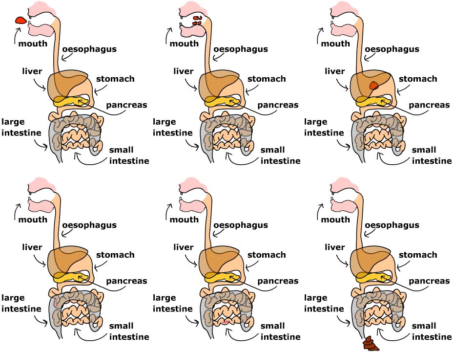
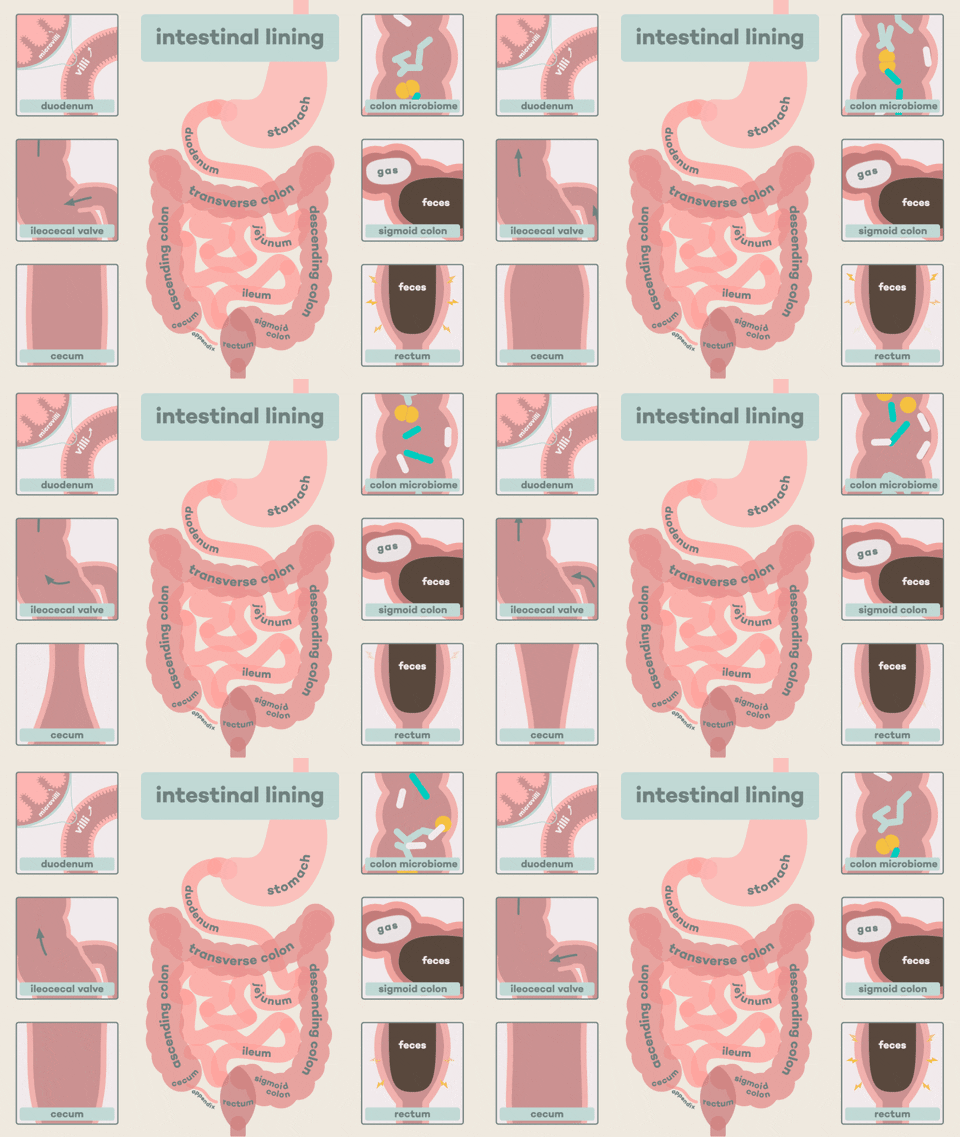
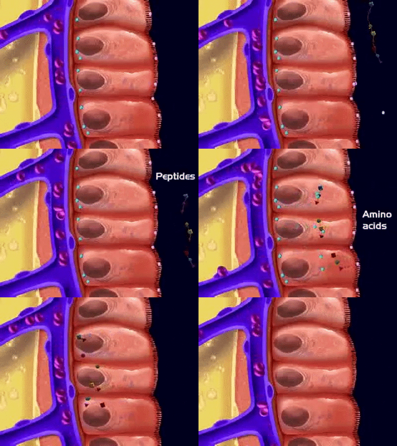
the EXTRAORDINARY tubular network of the small intestines and conclusion of the research project2/5/2021 The small intestine is the longest organ in the human body. It is the major site where food is broken down (digested) and the small soluble molecules, electrolytes and water are then absorbed into the systemic blood circulation with a minimal diffusion rate. The presence of microscopic projections called villi and microvilli in the intestinal wall increases the surface area of absorption for molecules to be transported to tissues and glands via the blood (OpenStax, 2021). Therefore, the small intestine is adapted for digestion and absorption because of the presence of enzymes, the regulation of gut hormones, motility, villi, and immunological factors for the microbiota homeostasis to ensure no harmful microorganisms such as bacteria enter the human body system (Campbell, 2016; Collins et al., 2020; Walters 2003). Besides, waste gases, for instance, carbon dioxide are removed and exhaled (Collins et al., 2020). Last month, we discussed the link between the anatomy and physiology of the musculoskeletal system with the Quran and Hadith. We also discussed how the bone and intestines are associated, particularly the microbiota. One of the molecules absorbed is the calcium ions and the malfunction of the bone has been found in lactose intolerance, coeliac disease, short bowel syndrome, bacterial overgrowth, and Crohn’s disease (Campbell, 2016; Augustyn et al., 2019). Therefore, understanding how the small intestine functions creates a framework of identifying pathways to treat gastrointestinal diseases (Volk et al. 2017; Zheng et al. 2020). Allah (The Most High) has revealed how the organs were nourished by the gastrointestinal system in the following verse: “Verily, in cattle there is a lesson for you. I give you drink from their insides, coming from a conjunction between the digested contents (of the intestines) and the blood, milk pure and pleasant for those who drink it.” [Quran, Surah Al Nahl (The Bee) 16:66] This highlights the role of enzymes in digesting food is primarily found in the mouth, stomach, pancreas, and small intestine. Ibn Kathir (2000) mentioned concerning this verse, that it is about one cattle or a herd of cattle, for we can provide a drink from their bellies. Also, the blood and milk have a defined route when the food is digested in the stomach i.e., the blood goes to the veins whereas the milk goes to the udder, the urine is excreted via the bladder and the faeces is excreted via the anus. The design of how Allah (The Most High) created the cattle is mirrored with what occurs in the human body where each defined route is not affected by the other. Subhanallah! For example, the difference in the pulmonary and systemic circulation in the human body where the former is involved with the lungs and the latter is involved to provide for body tissues. “So, which of the favors of your Lord would you deny?” [Quran, Surah Al Rahman (The Most Gracious) 55:13] The development of the small bowel in the foetus occurs in week 3; this is where gastrulation occurs. Gastrulation is where the tubular network and glands are formed in the primitive gut and consist of three germ layers: endoderm, mesoderm, and ectoderm (Moore, 1986). The process is regulated by the Hox signalling pathway primarily by the following genes: SOX9; FOXA2 and GATA4 (Volk et al., 2017). The midgut begins to open and associate with the disc of the embryo ca. week 4 (Fish and Burns, 2020; Moore, 1986). The lumen of the small intestine and the yolk sac form the vitelline duct. The development of the gut progresses over the weeks, the loop herniates on the umbilicus and this increases the surface area of the small intestine, large intestine, liver and kidneys to develop further (Collin et al., 2020). The epithelium undergoes various turnovers via stem cells. The endodermal layer forms the inner epithelial lining; the splanchnic mesoderm forms the muscular connective tissue, the jejunum and ileum derive from the midgut and the foregut from the duodenum (Collins et al., 2020). The cuboidal cells set up the basis for the villi to form (Collins et al., 2020). At week 10 of gestation, the small intestine moves to the abdomen and by week 11 the process is completed, and the crypt, villi and microvilli are produced (Fish and Burns, 2020). The neural crest cells produce the enteric nervous system by week 13 of gestation and the vagal nerve is the main nerve involved in the small intestine (Volk et al., 2017; Moore, 1986). Anatomy and Physiology of the small intestine.The small intestine is an important organ of the digestive system. This is where most of the food is digested, and nutrients are absorbed to allow the body tissues to undergo cellular and biochemical processes at a peak level. It is linked to the large intestine as presented in Figure 1 where the water and electrolytes are absorbed (Volk et al., 2017). The approximate length of the small bowel is 3 to 5 metres. It is divided into three sections: duodenum, jejunum, and ileum (Collins et al. 2020). This is ca. five times longer than the large intestine but smaller in diameter (Nigam et al., 2019). Volk et al. estimated the length of the small intestine is ca. 6 metres (Volk et al. 2017). This indicates that the mass and length of the small intestine takes a great portion of the abdominal cavity. The duodenum is the first and shortest section of the small bowel and is approximately 20 - 25 cm (Nigam et al., 2019). It is C-shaped where the proximal end of the duodenum is linked to the antrum of the stomach (pyloric sphincter) whereas the distal end is the start of the jejunum in the peritoneal cavity. The duodenum is further subdivided into superior, descending, horizontal and ascending duodenum (OpenStax, 2020). The superior sub-section is peritoneal, and the remainder of the duodenum is retroperitoneal (Collins et al., 2020). The chyme (partially digested food) that enters the duodenum is under acidic conditions due to the presence of hydrochloric acid in the stomach. It is mixed with enzymes from the pancreas, the bile from the liver, bicarbonate ions secreted by the duodenum, Brunner’s glands in the submucosa secretes an alkaline-based mucus that acts as a protective barrier against the acidic conditions (OpenStax, 2020; Collins et al., 2020). The presence of the ligament of Treitz comprises of skeletal muscle tissue that binds the duodenal-jejunal flexure. The bile duct and pancreatic duct join at the hepatopancreatic ampulla situated in the duodenal wall where it enters the duodenum through a tiny structure called the major duodenal papilla. The amount of bile and enzymes that enter the duodenum is regulated by the hepatopancreatic sphincter (sphincter of Oddi) (OpenStax, 2020). Figure 1: The digestive system The jejunum initiates at the duodenojejunal flexure and ends at the ileal section. It is distinctively different from the other sections of the small bowel due to the absence of Brunner's glands and Peyer's patches present in the duodenum and ileum, respectively. Its length is ca. 2.5 metres (Nigam et al., 2019). Other sources suggest the length is ca. 0.9 m (OpenStax, 2020). The primary function of the jejunum is the absorption of nutrients into the blood. Amongst the nutrients are glucose, fatty acids, glycerol, and amino acids. The jejunum is adapted for the absorptive function due to the presence of muscular flaps (plicae circulares), circular folds (Kercking valves), villi and microvilli. The Kercking valves are present near the mucosa and they flow in a spiral manner to increase the absorption rate. Microvilli are smaller finger-like projections than villi with an approximate length 1μm (0.001mm) and are present in the mucosal epithelial cells. They increase the surface area for absorption ca. 20-fold than villi (Fish and Burns, 2020). Villi are 0.5 – 1mm in length and have a capillary network comprising of an arteriole and venule where the latter allows glucose and amino acids to be absorbed directly into the blood whereas the fatty acids and glycerol are absorbed into the lymphatic vessels via the lacteals (Fish and Burns, 2020). The ileum is the longest section of the small intestine and is ca. 3 metres where it connects to the caecum of the large intestine via the ileocecal sphincter/valve as presented in Figure 2 (Collins et al., 2020). Other sources it is ca. 1.8 m in length and has a thicker vascular tissue with further mucosal folds (OpenStax, 2020). The function of the ileum is to absorb vitamin B12 and bile who were not absorbed in the duodenum nor jejunum - to be recycled (Collins et al., 2020). Another function of the ileum is to prevent the reflux of bacteria from the large intestine. The distal ileum has Peyer’s patches in the mucosa, a lymphoid tissue (lymphoid follicles) to prevent the bacteria from entering the blood (Fish and Burns, 2020). The number of Peyer's patches decreases with age (Fish and Burns, 2020). Besides, the dendritic cells in the mucosa sample the antigen from the blood, food, and microbiota. This modulates the activity of lymphocytes (CD8+ and CD4+) (Pederson et al., 2019). The ileal section is under hormonal and nerve control. Figure 2: The intestinal lining the layers of the small intestineThere are several layers in the small intestine: the inner layer is lined by mucosa responsible for the maximal absorption where enterocytes have the villi and crypt. The crypt cells are involved in proliferation and convert to enterocytes, Paneth cells, goblet cells and enteroendocrine cells (K and S) (Collin et al. 2020). The goblet cells secrete mucus in the intestinal glands and epithelium (OpenStax, 2020). Paneth cells secrete lysozymes that conduct a bactericidal action for macrophages to engulf them via phagocytosis (OpenStax, 2020). The MK and K cells secrete the insulinotropic peptide; a regulatory hormone that releases insulin. The S cells secrete secretin (OpenStax, 2020). Other cells found in the intestinal glands are G and I cell that secretes gastrin and cholecystokinin respectively. Cholecystokinin releases the pancreatic enzymes and bile. The M cells secrete motilin whose function is to increase the emptying of the stomach via peristaltic movement. It also stimulates the production of pepsin. The M cells are situated in the intestinal glands of duodenum and jejunum (OpenStax, 2020). The outermost layer is the serosa where it is subdivided into mesothelium and epithelium. These cells have a short life span of 3 to 5 days and therefore, has a high renewal rate (Collins et al., 2020). The middle layers of the small intestine are the muscularis and submucosa. The muscularis consists of smooth muscle, the outermost layer contains longitudinal muscle that shortens and elongates whereas; the innermost layer is a circular muscle that causes constriction. The presence of nerves between both muscle types increases the motility of the food from the proximal end to the distal end to conduct the chemical digestion (Collins et al. 2020). The submucosa is a connective tissue comprising of nerve, blood, and lymph supply (Collins et al., 2020; Hundt et al., 2020; Stallard et al., 1994). The nerve supply of the intestines arises from the autonomic nervous system. The vagus nerve of the parasympathetic system controls secretions and motility (OpenStax, 2020). The thoracic splanchnic nerve ganglion cells are part of the sympathetic system and surround the superior mesenteric artery (OpenStax, 2020). Pain stimulus is felt at this site. The main blood supply is via the superior mesenteric artery and vein. The arterial blood initiates from the celiac trunk to the superior mesenteric artery. The venous supply joins the splenic vein to form the hepatic portal vein and the blood containing the nutrients are transporter to the liver (Collin et al., 2020; OpenStax, 2020). The lymph supply via the nodes initiates from the mucosa to the mesentery to the arterial near the superior mesentery. The lymph then flows into cisterna chyli and then thoracic ducts and joins with the jugular and subclavian vein. It is the main pathway in how fat and immune cells are transported and how tumour cells can proliferate to other organs. This may explain why there is an enlarged node known as Virchow’s node that is clinically presented in small bowel tumours (Collin et al., 2020) An insight into the digestive and absorptive mechanisms of carbohydrateS Most of the chemical digestion takes place in the duodenum; this sub-section will briefly introduce how carbohydrates are digested. Carbohydrates are large polysaccharides - long chains of sugar units called monosaccharides joined together by glycosidic bonds; for instance, starch and glycogen. It is estimated that carbohydrates account to ca. 40-45% of our calorie intake and plant starches account for 50-60% of the diet (Goodman, 2010). Some of the food we eat contain disaccharides, for instance, maltose, lactose, and sucrose. Starch consists of two types of glucose polymers: a straight chain of glucose units linked together by alpha 1-4 glycosidic bonds called amylose and a branched form of glucose units linked together by alpha 1-6 called amylopectin. It functions as storage in plants. Following mechanical digestion by the teeth, the salivary glands secret an enzyme called alpha-amylase; it forms part of the saliva present the mouth. Enzymes have high specificity whereby a specific substrate binds to its active site just like a lock and key. The substrate that binds to amylase is starch - the product formed is maltose. Maltose is a disaccharide which consists of two glucose monomers connected through an alpha 1-4 glycosidic bond. It passes through the oesophagus (food pipe) to the stomach where chyme is produced. As the chyme enters the duodenum, the pancreas releases beta-amylase that chemically digest (hydrolyses) the disaccharides to produce the glucose molecules. Additional enzymes can be found in the brush border of the small intestine. Amongst them are sucrase, maltase, trehalase, isomaltase and beta-galactosidase. For instance, maltose can be hydrolysed by maltase through the alpha 1-4 glycosidic linkages. The product is glucose that is absorbed into the blood via the villi and is also transported by carrier-mediated transporters in the apical membrane (Fish and Burns, 2020). The two main transporters involved in transport are the facilitated diffusion transporter GLUT5 and Na+-coupled secondary active transport symporter (SGLT1). GLUT5 transports fructose and SGLT1 transports galactose and glucose. Glucose can also be carried by sodium ions (Goodman; 2010; Fish and Burns, 2020). An insight into the digestive and absorptive mechanisms of proteinS Proteins primarily function in growth and repair and the total daily protein is ca. 70 - 100 g of dietary protein (Goodman, 2010). They are long chains of amino acids linked together by peptide bonds. Proteolytic enzymes are required to hydrolyse the dietary protein into amino acids and smaller peptides. Some enzymes, for instance, endopeptidase breaks the internal bonds of polypeptides whereas, exopeptidases delete one amino acid at a time from the carboxyl group and the amino terminus of the protein. The chemical digestion of proteins initiates in the stomach where the active endopeptidase, pepsin, is secreted by chief cells in the gastric mucosa. It is activated from the zymogen pepsinogen and begins to digest proteins to produce the smaller polypeptides. Approximately, ca. 10 - 15% of proteins are digested in the stomach (Goodman, 2010). Pepsin differs from other enzymes because, despite its protein nature, it functions under acidic conditions because of the hydrochloric acid. Hydrochloric acid not only kills bacteria and other microorganisms but facilitates digestion by denaturing the proteins causing unfolding (Fish and Burns, 2020; Goodman, 2010). As the chyme enters the duodenum, the pancreas releases protease enzyme (under the control of cholecystokinin) and bicarbonate through the hepatopancreatic sphincter. Bicarbonate neutralises the hydrochloric acid that increases the pH to the optimum to elevate enzyme activity. Additional enzymes are secreted in the intestines: endopeptidases (elastase, trypsin, chymotrypsin) and carboxypeptidases A and B to further hydrolyse the polypeptides to 30% free amino acids and 70% of oligopeptides (Goodman, 2010). Oligopeptides consist of 2 - 8 amino acids and are further cleaved at its amino terminus by exopeptidases to produce dipeptides, tripeptides, and free amino acids. Elastase cleaves elastin and cleaves peptide bonds adjacent to alanine, serine, and glycine. Trypsin is activated from its zymogen trypsinogen by the jejunal brush border enteropeptidase/enterokinase. Trypsin also cleaves peptide bonds of other zymogens near the arginine or lysine amino acid residues to become active enzymes. On the other hand, chymotrypsin hydrolyses peptide bonds adjacent to hydrophobic amino acids - less specific (Goodman, 2010). Carboxypeptidases cleaves the carboxyl terminus of the polypeptides and have two types that specifically releases amino acids. Carboxypeptidase A releases alanine, leucine, valine, and isoleucine whereas, carboxypeptidase B releases lysine and arginine residues. The uptake of amino acids, tripeptides and dipeptides across the enterocytes requires carrier-mediated active and facilitated transport proteins as presented in Figure 3. The type of transporter is dependent on several factors: the type of amino acid and mechanism used for transport. There are various types: amino acids: anionic (acidic), neutral, cationic (basic), and zwitterion (Goodman, 2010) If the amino acid transporter has stereospecificity, they transport peptides with L-amino acids that have a greater affinity to bind than D-amino acids. They are actively co-transported with sodium ions via the capillaries of the villi to the liver via the hepatic portal vein (Goodman, 2010). Neutral L-amino acids are absorbed across epithelial cells by entering via the secondary active Na+-dependent cotransporter and leaving with the Na+-independent facilitated diffusion transporter. Other amino acids have a broad specificity whereby some undergo active transport whereas other transport via facilitated diffusion and can bind to different types of transporters across the basolateral membrane into the blood. Oligopeptides, most dipeptides, and tripeptides are predominantly transported by PEPT1 intestinal oligopeptide transporter residing in the duodenum and jejunum. PEPT1 has a low affinity and requires a hydrogen ion electrochemical gradient that initiates from the lumen of the small bowel to the cytosol of enterocytes. This is maintained by the Na+/H+ exchange in the brush border membrane removing the sodium ions by Na+, K+-ATPases across the basolateral membrane. This explains the difference in pH between the intracellular pH of the enterocyte 7.0 – 7.2 and the pH of the brush border 6.0. The deficiency of transporters or loss of function in kidneys can cause amino acids in the urine (Goodman, 2010). Figure 3: The absorption of peptides An insight into the digestive and absorptive mechanisms of fatS The products of the chemical digestion of lipids are fatty acids and glycerol. Lipids are initially digested in the stomach where gastric lipase cleaves 15-20% of fats and is completed by the pancreatic lipase in the duodenum (Goodman, 2010). Gastric lipase is secreted by the gastric chief cells in the fundic mucosa in the stomach. It has a pH of 3 - 6 and hydrolyses the ester bond to produce diglyceride and fatty acids. Cholecystokinin stimulates the emptying of the stomach that gives time for the lipases to digest the fats. The gallbladder is contracted to cause the hepatopancreatic sphincter to relax. The bile and pancreatic lipase are then released through the sphincter. Additional pancreatic lipases are colipase, cholesterol ester hydrolase and phospholipase A2. The bile salts can inactivate the lipase and the pancreatic colipase counteracts this. Colipase is activated by trypsin in the jejunum and binds to triglyceride (fat) that facilitates the binding between lipase and triglyceride. The bile salts emulsify fats from large droplets to small droplets and the reduction in the surface area of the fats facilitates the pancreatic lipase. The droplets convert into several forms by the addition of bile salts before producing the products: free fatty acids and 2-monoglyceride. At first, multilamellar liquid crystals are formed and are converted into unilamellar vesicles then mixed micelles (cholesterol, monoglycerides, bile salts, fatty acids, lysophospholipids) and then the final product (Fish and Burns, 2020; OpenStax, 2020; Goodman, 2010). Fatty acids are transported across the endoplasmic reticular brush-border membranes of the enterocytes and are absorbed into the blood. They are coated with lipoproteins to form chylomicrons that are taken via the central lateral of the villi to the lymph then the thoracic duct and the blood. An example of a chylomicron is the apoprotein B48 - a large, hydrophobic protein and like other chylomicrons, it is dependent on the microsomal triglycerides transport protein (MTP) binds with a dense particle to form a complex and catalyses the transfer from a triglyceride to the droplet (Goodman, 2010). A pre-chylomicron is formed between the Apoprotein B48-dense particle complex and the apoprotein AIV from the smooth endoplasmic reticulum. It is then transported to the Golgi apparatus via a specific transport vesicle to complete the process. Chylomicrons are transferred by another vesicle to the basolateral membrane to be exocytosed into the lacteal lymph vessel (Lowe, 2002; Goodman, 2010). Mixed micelles depend on the electrochemical gradient where the Na+/H+ exchange cause the water-soluble (hydrophilic) fatty acids to protonate and release. They are then bound to serum albumin and absorbed directly into the portal blood. Moreover, fatty acid translocase (FAT/CD36) mainly expressed in the proximal intestinal mucosa uptakes long chains of fatty acids into the enterocytes (Lowe, 2002). Carboxyl ester hydrolase cleaves dietary cholesterol. The uptake of cholesterol is conducted by Neimann-Pick C1-like 1 (NPC1L1). Efflux transport is conducted by ATP-binding cassette proteins (ABCG5 and ABCG8) (Lowe, 2002). Malabsorption of fats can influence the structure and function of the whole mucosa. This can lead to oily stools (steatorrhea) that are clinically presented in patients with pancreatic insufficiency and coeliac disease and may also affect the absorption of vitamin K, E, D and A. For instance, Vitamin D requires chylomicrons to be absorbed, other examples are diarrhoea and milk intolerance (Burns and Fish, 2020; Nikaki and Gupte, 2017). An insight into the digestive and absorptive mechanisms of vitamins and minerals. The vitamins vary where they are absorbed, vitamin K, A and D are lipid-soluble (lipophilic) and are absorbed passively. Vitamin B, however, is a water-soluble protein and is required for erythropoiesis, hair, nails and the contraction of muscles and conduction of nerve impulses to maintain the activity of the heart and brain. Vitamin B9 (folate) is absorbed in duodenum and jejunum (Fish and Burns 2020). Vitamin B12 binds to R protein in acid milieus of the stomach and then dissociates from the complex when reaching the duodenum. B12 (cobalamin) binds to the intrinsic factor secreted by gastric parietal cells before being absorbed into the terminal ileum and enter the enterocyte via ileal membrane receptors. It is required for the synthesis of DNA, red blood cells, metabolic process, and nerves. They are then absorbed into the blood by associating with transcobalamin II (Nigam et al., 2019). A B12 deficiency can destroy the parietal cells by autoantibodies which influences and decreases the production of hydrochloric acid. This leads to pernicious anaemia and even dementia if untreated (Nigam et al. 2019). The movement towards the large intestineFollowing absorption, the small bowel becomes less distended, and the gastroileal reflex stimulates peristalsis to move the remaining unabsorbed contents from the ileum to the caecum of the large intestine. With each motility action, the ileocaecal valve relaxes. This ensures that the meal eaten is completely emptied from the stomach and small bowel before the next meal. It takes ca. five hours for contents to leave the small intestine (Nigam et al., 2019; Hundt et al., 2020). Allah (The Most High) states regarding the large intestine: "Is the description of Paradise, which the righteous are promised, wherein are rivers of water unaltered, rivers of milk the taste of which never changes, rivers of wine delicious to those who drink, and rivers of purified honey, in which they will have from all [kinds of] fruits and forgiveness from their Lord, like [that of] those who abide eternally in the Fire and are given to drink scalding water that will sever their intestines?" [Quran, Surah Muhammad, 47:15] This complements the findings by researchers whereby boiling water can affect the peritoneum and the outer layer of the intestine where there are Pacini bodies containing nerve endings and will feel the pain. The intestines have no receptors to sense the stimulus (Al Ghazal, 2006). The large intestine absorbs water containing mineral ions and nutrients released by the bacteria and is used for metabolic processes. This solidifies the condensed faeces and is stored in the sigmoid colon and rectum where it is excreted via the anus in a process called defecation. The evidence of hadith on the intestinesThere are several hadiths in which our Beloved Prophet (peace be upon him) refers to the intestines. One of which is the following: It was narrated by Abu Hurairah (may Allah have mercy upon him) that the Prophet Muhammad (peace be upon him) said: "A man used to eat much, but when he embraced Islam, he started eating less. That was mentioned to the Prophet who then said, "A believer eats in one intestine (is satisfied with a little food) and a Kafir eats in seven intestines (eats much)." [Al-Bukhari, Book 65, Hadith 308;309] One of the respected scholars, Al Nawawi reported that the most agreed opinion is some believers eat one intestine and most non-believers eat seven intestines. It does not necessitate that each one of the seven intestines is like that of the believer. The hadith emphasizes that one needs to be content with little from this temporary world and eating a lot can have a negative effect and the opposite. This also links with the following verse from the Quran where Allah (The Most High) states: “O children of Adam, take your adornment at every Masjid, and eat and drink, but be not excessive. Indeed, He likes not those who commit excess.” [Quran, Surah Al-Araf (The Heights); 7:31] Regarding this verse, Al Sarakhsi mentioned in his book Al-Mabsoot: "That is because one eats to benefit himself; so, there is no benefit in eating more than one's fill for it causes harm. Accordingly, this will be like throwing food in garbage or worse than that. Also, other people have a right in the food that is more than one's need for it may stratify another person's hunger if it reaches him with or without a return. Thus, eating it involves a violation of the right of others and this is something prohibited. Moreover, eating more than one's fill may cause diseases and thus this will be a form of hurting oneself." Therefore, it is important to maintain a balanced diet where Prophet (peace be upon him) said: "The son of Adam cannot fill a vessel worse than his stomach, as it is enough for him to take a few bites to straighten his back. If he cannot do it, then he may fill it with a third of his food, a third of his drink, and a third of his breath." [Al-Nasai 4:178, Al-Tirmidhi, Ahmad 4:132; Tuhfat Al Ahwadhi 7:51] Tumours of the small intestineSmall intestinal cancers are rare and around 1,700 people are diagnosed annually between the period 2015 - 2017 where ca. 790 in females in 2017 and ca. 970 in males in 2017 (Cancer Research UK, 2021). It is prevalent amongst people aged 80 to 84. The use of techniques, such as molecular profiling and enteroscopy allows tumours to be diagnosed early leading to therapeutic modalities. However, the survival rate remains the same and this is because of the lag time. However, the use of cancer waiting times allowed tumours to be diagnosed most quickly - progress (Islam et al., 2014; Pedersen et al., 2019). The main type of small intestinal cancer is the small bowel adenocarcinoma (Pedersen et al., 2019). This research area has increased because many previous studies observed how colorectal cancer (tumours of the large bowel) can metastases to this defined region which allows it to be resectable. The cause of this rare tumour is due to several respects: firstly, the dilute food contents have fewer effects on the duodenal mucosa than the large intestine. There is also a difference in the redox microenvironment where there are less reactive oxygen species (ROS) in the small intestine than the large intestine. Therefore, DNA adducts are readily formed in the large intestine and this causes DNA damage, and this influences the immunological control than the small intestine. This occurs despite dendritic cells are present in the mucosa and the release of interleukin 10 against the cell transformation. The levels of ROS can increase due to several risk factors: smoking cigarettes and alcohol (Pedersen et al., 2019). The role of microbiota homeostasis in promoting or suppressing tumorigenesis and proliferation in the small intestines is still ongoing (Pedersen et al. 2019). Patients with commodity conditions i.e., coeliac disease and Crohn's disease are also at risk. Other causes of small bowel cancer could be genetically linked where germline mutations in genes involved in DNA mismatch repair MLH1, MSH2, MSH6, PMS2 have been revealed and may also affect colorectal cancers that require immune checkpoint therapy. Moreover, patients with familial adenomatous polyposis that occurs in the early the onset of colorectal tumours have a 4.5% chance of developing small bowel adenocarcinoma (Pedersen et al., 2019). The clinical presentation of small bowel adenocarcinoma is normally intermittent abdominal pain. Other symptoms include nausea, weight loss and gastrointestinal bleeding, however, the latter can be misinterpreted as iron deficiency anaemia because of the negative outcome of the colonoscopy test (Pedersen et al., 2019). The rarity of this cancer did not allow a screening programme to be established like the NHS large bowel screening programme. However, it can be diagnosed through radiological imaging to determine whether it has progressed from the colon. CONCLUSION OF RESEARCH PROJECTOverall, we have reached the end of my six/seven-month 40,803-word literature review (excluding references and tables) titled: 'The link between medicine and science with the Glorious Quran... with a focus on anatomy and physiology'. It was divided into six sections:
The two foundations of Islam are Quran and Hadith that teach us to respect, learn tolerance of other religions, and persuade us to be successful in medicine and science. The Greek medical and scientific writing was translated to Arabic in Syria, Egypt and Iraq. This led to Arabic become a language of not only the Quran but a language of effective diplomacy, learning and long-lasting innovations. The Quran has been written in the traditional language of Arabic. It comprises of 114 chapters and 6237 verses distributed to Prophet Muhammad (peace be upon him) over 23 years which comprised of 13 years in Makkah and 10 years in Madinah. The Quran consists of parables of the Prophet, divine nature, social justice, and lessons that we can learn and apply once a thorough understanding is gained because some verses can be misinterpreted and it is through the scholars of exegesis (Tafsir) one can thoroughly understand what is meant by what is said (Ibn Kathir, 1990). This provides feelings of peace, calmness, patience, and tranquillity. Naturally, the pandemic can cause upset but through reading the Quran one can counteract this. Allah (The Most High) states: “There is no god but He: That is the witness of Allah, His angels, and those endued with knowledge, standing firm on justice. There is no god but He, the Exalted in Power, the Wise” [Quran, Surah Al Imran (Family of Imran) 3:18]. There were several questions asked by readers and I wanted to leave it till the end. One of the lessons I learnt from Surah Kahf, is when Prophet Moses (peace be upon him) accompanied Khidr on his journey and the Prophet wanted to find out the reason why Khidr did a particular action for each event and each time, Khidr replied '“Did I not say that with me, you would never be able to have patience?”' [Quran, Surah Kahf, 73:74] First question: What made you start this project? Like everyone else, I was also affected by the coronavirus pandemic and I tried to respond to it in a positive mindset by increasing my relationship with Allah (The Most High) through the Quran more than a normal day, being surrounded by my loved ones and my zeal for learning new things. I wanted to combine my interests in medicine, research and the Quran. We can easily count the effects of the pandemic, however, the blessings we have within us and around us are countless. The engineering mechanisms of how our bodies function have been designed and created by Allah (The Most High) - no one can ever reach its status. How have we existed? How cells interact with one another? How are nutrients circulated? How is movement conducted? The visual concepts and other senses that involve emotion, contemplation and building of our inner faith? As a human and Muslim, I must be conscious of my meaning and purpose - and so do you. The anatomy and physiology of the human body are a piece of living evidence. It illustrates the intellectual honesty of our limits as human beings and what we know. I have spent months in my spare time reading, understanding, revising, and learning new knowledge through research - this is only a drop of the ocean. There is so much more to learn, so much more to write - seeking knowledge is part of my worship. This is merely an introduction between both fields of medicine and Tafseer. The respected scholars previous and current have studied each verse or surah or even understanding the hadith for years and continue to learn and develop. And despite their experience and present level of their knowledge, there are still aspects in the Quran and Hadith that are incomprehensible. “And those who strive in Our (cause), – We will certainly guide them to our Paths: For verily Allah is with those who do right” [Quran, Surah Al Ankabut (The Spider) 29:69]. Second question: Why is it published at 8 pm UK and not earlier? I thought that whilst searching for a job, I aimed to ensure that this project would not influence my work and family commitments. For instance, not publishing on time, so I thought publishing it in the evening will prevent this limitation inshaAllah (if Allah wills). Having a positive mindset can help with any trial, such as this pandemic. We have a duty of care to learn, ask questions and be better through observation, reasoning and productivity. It was narrated by Anas ibn Malik (may Allah have mercy upon him): “Seeking knowledge is an obligation upon every Muslim.” [Sunan Ibn Majah, 224] I thought I would continue with the planned time and share what I read and learnt. The first verse revealed to our Beloved Prophet (peace be upon him) on the 27th of Ramadan 611 AD is the following: “Proclaim! (or read!) in the name of thy Lord and Cherisher, Who created – Created man, out of a (mere) clot of congealed blood: Proclaim! And thy Lord is Most Bountiful, He Who taught (the use of) the pen, Taught man that which he knew not” [Quran, Surah Al Alaq (The Clot) 96: 1-5]. Being able to read and devote ourselves to the pursuit of seeking knowledge can protect our faith for Allah (The Most High) states: “And it is not for the believers to go forth [to battle] all at once. For there should separate from every division of them a group [remaining] to obtain understanding in the religion and warn their people when they return to them that they might be cautious." [Quran, Surah Al-Tawbah (Repentance) 9:122]. Third question: Why have you mentioned the Quran and not the hadith on the poster? I wanted to begin with Allah (The Most High) - words of the Almighty who created me and you. The main source of our life and what we recite in our daily prayers. Verily, without doubt, the Quran and Hadith are both important. The authentic hadith are collections of sayings by our Beloved Prophet (peace be upon him) by respected narrators where logic and reason with authenticity are combined and graded as Sahih. The overall evaluation and classification are known as the Mustalahul Hadith, this can prevent conflicts of principles and revelations (Hasan, 1994; Al Suyuti, 2017; Ismail et al., 2014; Moore, 1986). Similarly, as the understanding of the scholars in Tafseer and Hadith are increasing through various in-depth research, understanding the human body is also constantly evolving through various research methodologies and techniques to understand the cellular and biochemical mechanisms that affect the human body in normal physiological and pathophysiological consensus. Clinical research is fundamental in healthcare, where addressing medical needs is met through advancing knowledge to discover better treatment and procedures to improve patient outcomes. Ultimately, our physical growth may stop at puberty, but our mental and emotional growth has no end. Reading, listening, understanding and other communication skills are survival mechanisms that create new cumulative dimensions to learn something new every day no matter how new or how simple the information is (Syed, 2006). This is not limited to textbooks or articles but also involves collaborative interactions. I hope you enjoyed reading my literature review just like how I enjoyed the whole process and what you learn and what I learn is all part of our growth. So high [above all] is Allah, the Sovereign, the Truth. And, [O Muhammad], do not hasten with [recitation of] the Qur'an before its revelation is completed to you, and say, "My Lord, increase me in knowledge." [Quran, Surah Taha 20:114] referencesAl-Ghazal, S. (2006) Medical Miracles of the Qur'an UK: Islamic Foundation
Al Sarakhsi (1986) Al Kitab Al Mabsut Beirut: Dar Al-Kotob Al-Ilmiyah Al-Suyuti, A. (2017) Tadrib al-Rawi fi Sharh Taqrib al-Nawawi. Cairo: Dar Ibn Al-Jawzi. Augustyn, M., Grys, I. and Kukla, M., (2019) Small intestinal bacterial overgrowth and nonalcoholic fatty liver disease. Clinical and Experimental Hepatology, 5(1), pp.1-10. Campbell, J., Berry, J. and Liang, Y., (2019) Anatomy and Physiology of the Small Intestine. Shackelford's Surgery of the Alimentary Tract, 2 Volume Set, pp.817-841. Cancer Research UK (2021) Small intestine cancer statistics. Available [online] https://www.cancerresearchuk.org/health-professional/cancer-statistics/statistics-by-cancer-type/small-intestine-cancer#heading-Zero Collins, J.T., Nguyen, A. and Badireddy, M. (2020) Anatomy, Abdomen and Pelvis, Small Intestine. Available [online] https://www.ncbi.nlm.nih.gov/books/NBK459366/ Fish, E.M. and Burns, B. (2020) Physiology, Small Bowel. Available [online] https://www.ncbi.nlm.nih.gov/books/NBK532263/ Goodman, B., (2010) Insights into digestion and absorption of major nutrients in humans. Advances in Physiology Education, 34(2), pp.44-53. Hasan, S. (1994) An introduction to the science of hadith. London: Al-Qur’an Society. Hundt, M., Wu, C.Y. and Young, M. (2020) Anatomy, Abdomen and Pelvis, Biliary Ducts. Available [online] https://www.statpearls.com/articlelibrary/viewarticle/32108/ Ibn Kathir, I. (1990) Tafsir Al-Qur’an al- ‘Adheem. Al-Zarqa: Maktabah Al-Manar. Ibn Kathir (2000) Tafsir Ibn Kathir. Saudi Arabia: Darussalam. Islam, R., Leighton, J. and Pasha, S., (2014) Evaluation and management of small-bowel tumors in the era of deep enteroscopy. Gastrointestinal Endoscopy, 79(5), pp.732-740. Ismail, T.M.S.T., Baru, R., Hassan, A.F., Salleh, A.Z.B. and Amin, M.F.M. (2014) The matan and sanad criticisms in evaluating the hadith. Asian Social Science; 10:152. Kiela, P. and Ghishan, F., (2016) Physiology of Intestinal Absorption and Secretion. Best Practice & Research Clinical Gastroenterology, 30(2), pp.145-159. Ma, Z. and Lee, Y., 2020. Small intestine anatomy and physiology. Clinical and Basic Neurogastroenterology and Motility, pp.101-111. Moore, K.L. (1986) A scientist's interpretation of references to embryology in the Qur’an. Journal of the Islamic Medical Association of North America 18:15-17. Nigam, Y., Knight, J. and Williams, N. (2019) Gastrointestinal tract 4: anatomy and role of the jejunum and ileum. Nursing Times [online]; 115: 9, 43-46. Nikaki, K. and Gupte, G., (2016) Assessment of intestinal malabsorption. Best Practice & Research Clinical Gastroenterology, 30(2), pp.225-235. OpenStax (2021) 23.5 The Small and Large Intestines Available [online] https://openstax.org/books/anatomy-and-physiology/pages/23-5-the-small-and-large-intestines Pedersen, K., Raghav, K. and Overman, M., (2019) Small Bowel Adenocarcinoma: Etiology, Presentation, and Molecular Alterations. Journal of the National Comprehensive Cancer Network, 17(9), pp.1135-1141. Stallard, D., Tu, R., Gould, M., Pozniak, M. and Pettersen, J., (1994) Minor vascular anatomy of the abdomen and pelvis: a CT atlas. RadioGraphics, 14(3), pp.493-513. Syed, I. (2006) ‘The pleasures of seeking knowledge’ Available [online] https://www.islamawareness.net/Knowledge/knowledge_article0003.html Volk, N. and Lacy, B., (2017) Anatomy and Physiology of the Small Bowel. Gastrointestinal Endoscopy Clinics of North America, 27(1), pp.1-13. Yusof, N., Che Mohamad, C. and Hassan, A., (2018) Anatomy of Musculoskeletal System in the Light of the Qur’an and Hadith. IIUM Medical Journal Malaysia, 17(1).
0 Comments
|
Archives
February 2021
Categories
All
|
© COPYRIGHT 2013. ALL RIGHTS RESERVED.
Proudly powered by Weebly
 RSS Feed